Serous atrophy of bone marrow


Clinical History
A 33-year-old woman was admitted to our department because of pain in right knee.
Her medical history was characterized by anorexia nervosa (AN) and personality disorder type AN binge purging. Her BMI was of 11.9 kg/m2.
Previously known substance and alcohol abuse in adolescence.
Imaging Findings
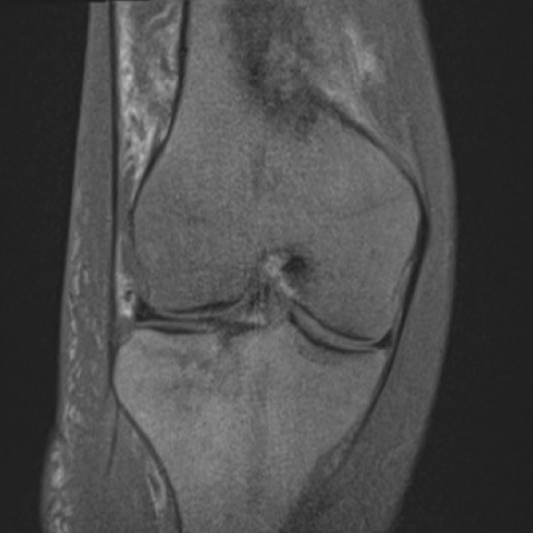
MRI of the knee showed severe changes in the intra-medullary fat with mildly low signal intensity in T1 weighted sequence and high signal intensity in STIR, which raised the suspicion of a technical problem.
For this reason, also a DP FS sequence was performed, confirming similar signal intensity and no fat signal suppression.
These findings are typical of the so-called “Flip-Flop effect”: it refers to a confusing MRI signal of the skeletal system that occurs in clinical conditions in which a severe fat depletion is present.
Similar findings were observed at the level of the subcutaneous tissues, with high signal intensity on all fat-suppressed fluid-sensitive sequences.
Coronal T1-weighted image showed some hypointense lines in the lateral tibial plateau, parallel to the joint surface, in keeping with insufficiency fractures.
The presence of a fair amount of joint effusion was also noted.
Discussion
Serous atrophy of bone marrow, also known as gelatinous transformation of bone marrow, is a non-neoplastic disease that occurs with chronic illness and poor nutritional status with weight loss. Anorexia nervosa, malabsorption, chronic infections (e.g. AIDS, tuberculosis), malignant tumours, chronic heart failure, chronic kidney disease, alcoholism, cytotoxic drugs [1], acute febrile illness and myelodysplastic syndrome have been described as underlying conditions. Scientific literature has always shown a considerable interest in the connection between serous atrophy and anorexia nervosa [2].
From a clinical point of view, severe weight loss and anaemia are typical [3], however, a general decrease in hematopoiesis can lead to leukopenia, with increased risk of infection [4]. The major complication is the increased risk of fractures, due to the impairment of bone quality [5].
Serous atrophy of bone marrow is due to a massive fat mobilisation – in case of prolonged negative metabolic balance. A material made of hyaluronic acid-rich musopolysaccaride with jelly-like consistency fills the extracellular space in the cancellous bone. This criterion, together with a decrease in size and number of fat and hematopoietic components, is essential for the histopathological diagnosis, that takes place after the biopsy [3].
The term “Flip-Flop effect” derives from the inverse signal characteristics of medullary fat inside the bone. It is used in literature to characterise this disease.
The T1 weighted images seem to be fat-suppressed, with mildly low signal intensity of the bone marrow. The images sensitive to the suppressed fluid-inversion recovery as well as T2 with chemical fat suppression - appears to be non-fat-suppressed with high signal [6].
Fat depletion is also visible on MRI (decrease in volume of fat anywhere where there is fatty tissue), and lipodystrophy is characterized by decreased signal intensity on T1, increased on T2 and water-sensitive sequences and increased attenuation of the fat on CT.
These features can cause misdiagnosis: this type of signal could be mistaken for a technically defective MRI study.
The marrow does not enhance after paramagnetic contrast administration. This helps in the differential diagnosis with diffuse malignancy [7]. Chronic osteomyelitis may also be considered in the differential diagnosis for the impairment of bone quality, but the presence of cortical destructive changes, intramedullary abscess formation, cloaca formation, and, soft tissue inflammation, can help differentiate.
Take-home points
- Serous atrophy is a non-neoplastic disease of bone marrow that occurs with chronic disease, mostly anorexia nervosa.
- The major complication is the increased risk of insufficiency fractures due to the bone quality impairment.
On MRI, the marrow has mildly signal intensity on T1 weighted sequences, while it has high signal intensity on images sensitive to the suppressed fluid – Flip-Flop effect.
Differential Diagnosis List
Final Diagnosis
Serous atrophy of bone marrow
Liscense
This work is licensed under a Creative Commons Attribution-NonCommercial-ShareAlike 4.0 International License.
Figures


Medical Analysis Report
I. Radiological Findings
The MRI of the patient’s right knee shows:
- In T1-weighted images, the bone marrow signal of the distal femur and proximal tibia is generally reduced, presenting a mild diffuse low signal.
- In T2-weighted images and other water-sensitive sequences (such as STIR or FS T2), the signal in the above-mentioned bone marrow regions is relatively increased, locally showing a “Flip-Flop effect” (indicating that the fat signal appears suppressed in T1-weighted images while showing relatively high signal intensity in T2-weighted images).
- The overall contour of the articular cartilage remains acceptable, with no obvious localized cartilage defects or large-scale destruction. No significant narrowing of the joint space is observed.
- No obvious disruption of trabecular structure or cortical bone continuity is observed, and there is no clear evidence of fracture lines.
- No significant swelling, effusion, or abscess in the periarticular soft tissue is detected, and no obvious erosion or destruction is noted.
Overall, these characteristics suggest decreased fatty components of the bone marrow. Considering the patient’s clinical history, serious nutritional deficiency or physical depletion-related bone marrow changes should be highly suspected.
II. Potential Diagnoses
- Serous Atrophy of Bone Marrow
Associated with chronic malnutrition, severe weight loss, or a prolonged negative energy balance. The patient has severe anorexia (BMI as low as 11.9 kg/m2), consistent with a typical etiology. The “Flip-Flop” signal changes on imaging match the extensive loss of bone marrow fat. - Diffuse Malignant Infiltration of the Bone Marrow
Conditions like lymphoma or metastatic tumors can also cause low signal on T1-weighted images and high signal on T2 or water-sensitive sequences. However, such diseases are usually accompanied by other laboratory or clinical features (abnormal blood work, systemic symptoms, etc.), and contrast enhancement often shows varying degrees of enhancement. - Chronic Osteomyelitis/Bone Infection
Chronic infection can lead to bone marrow signal abnormalities, but typically presents with focal lesions in the medullary cavity, soft tissue edema, or destructive changes, which are not completely consistent with this patient’s findings.
III. Final Diagnosis
Considering the patient’s severe anorexia history (AN, with marked underweight and malnutrition) and the typical “Flip-Flop” appearance on imaging reflecting depletion of bone marrow fat, the most likely diagnosis is:
Serous Atrophy of Bone Marrow.
It is recommended to supplement with further laboratory tests (such as blood tests, bone density assessments, nutritional indicators) and consider bone marrow biopsy to rule out diffuse malignant bone marrow infiltration or other rare pathologies. If definitive clarification is required, histological diagnosis via biopsy is advised.
IV. Treatment and Rehabilitation Plan
Based on the above diagnosis and the patient’s history, the main treatment and rehabilitation recommendations include:
1. Nutrition and Medical Treatment
- Develop a reasonable nutritional supplementation and gradual weight gain plan under the guidance of professional nutrition and psychiatry teams, correcting electrolyte imbalances and other deficiencies.
- Monitor bone density and hematological indicators. If necessary, provide calcium supplementation, active vitamin D, or anti-osteoporosis medications.
- For coexisting personality disorders, emotional problems, or potential substance dependence, incorporate psychotherapy and specialized interventions.
2. Exercise Prescription and Rehabilitation Training
Once nutritional status improves and body weight increases, a gradual rehabilitation and exercise routine should be implemented as follows:
- Initial Stage (Recovery Phase):
• Goal: Improve cardiopulmonary function, enhance basic muscle strength and body coordination.
• Exercise forms: Low-impact activities such as seated cycling, simple bodyweight exercises (e.g., light resistance lower limb extensions, core stability training), 3-4 times per week, about 20-30 minutes each session.
• Intensity: Primarily low to moderate; ensure the ability to speak comfortably during exercise. - Intermediate Stage:
• Goal: Gradually increase bone loading to promote bone regeneration and improve bone density, enhance muscle endurance.
• Exercise forms: Under professional supervision, gradually transition to strength training (light to moderate resistance exercises, elastic band or small dumbbells) and low-impact treadmill brisk walking/jogging 3-4 times per week, 30-45 minutes each session.
• Intensity: Maintain moderate intensity; monitor heart rate and blood pressure; avoid excessive fatigue. - Advanced Stage:
• Goal: Once bone quality and muscle function are stable, enhance daily activity capabilities and overall fitness.
• Exercise forms: Gradually introduce light fitness running and non-collision ball sports (such as table tennis or badminton), or engage in more systematic resistance training under professional coaching.
• Intensity and Frequency: Increase gradually according to fitness levels, 3-5 times per week.
Throughout rehabilitation, close attention should be paid to skeletal and joint status, as the patient’s bones are more fragile. Avoid sudden, high-impact, or high-intensity exercises. Ensure full warm-up before exercise and adequate stretching and relaxation afterward.
Disclaimer:
This report is based on the provided patient information and imaging data for analysis and is for reference only. It is not a substitute for in-person diagnosis and the guidance of professional physicians. If you have any questions or notice any changes in symptoms, please seek medical attention or consult a specialist promptly.
Human Doctor Final Diagnosis
Serous atrophy of bone marrow