Sinding-Larsen and Johansson Syndrome: MRI findings


Clinical History
14-year-old presented with pain in anterior knee over 5 months time without any history of direct trauma.
Imaging Findings
14-year-old presented with pain in anterior knee over 5 months time without any history of direct trauma. Pain is worse during sporting activities. On examination patient is tender over inferior part of patella and over patellar ligament. MRI scan was done to confirm the clinical diagnosis and to rule out other knee pathologies.
Discussion
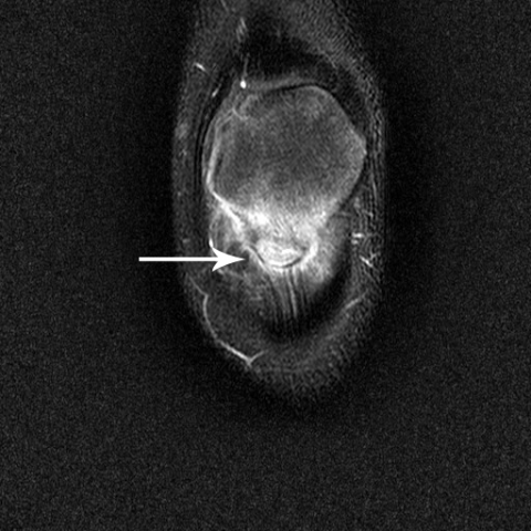
Sinding-Larsen and Johansson syndrome was described independently by Norwegian physician Christian Magnus Falsen Sinding-Larsen and Swedish surgeon Sven Christian Johansson. It is a type of osteochondrosis at the proximal attachment of patellar ligament, which is common in adolescents especially those who participate in jumping activities. The aetiology appears to be traction tendinitis with de novo calcification in the proximal attachment of patellar tendon [1]. This may be caused by repetitive microtrauma to the tendon at the insertion to lower pole of patella. Clinically, patients present with pain, swelling and tenderness over inferior pole of patella precipitated by overstretching or trauma. Although it’s mainly a clinical diagnosis, imaging modalities help in diagnosis. Plain x-ray imaging may show osseous fragmented appearance of lower pole of patella [1]. MRI findings include fragmentation of lower pole of patella, marrow oedema within the fragments, thickening of patellar tendon at its insertion and oedema of Hoffa’s fat pad. Although ultrasound imaging is also effective and can be used for periodic follow-up during the course of this condition, the advantage of MRI is that it allows ruling out other intra-articular derangements [2-4]. As it is self-limiting condition, treatment involves conservative methods. The condition can last from three to twelve months [1]. Restricting activities such as jumping, kneeling, squatting, stair climbing and running should be avoided during the course of this condition. In severe cases brief period of immobilisation of affected knee joint may be beneficial. In the end the fragmented lower pole of patella may get incorporated into patella.
Differential Diagnosis List
Final Diagnosis
Sinding-Larsen and Johansson Syndrome
Liscense
Figures
Sagittal T1 weighted Image of Knee

Sagittal T2 weighted image of Knee

Coronal T2 weighted image of Patella
1. Imaging Findings
Based on the provided MRI images of the knee, the following observations are noted:
• There is a lobulated or fragment-like signal change at the inferior pole of the patella, with local signs of calcification or bony fragments, suggesting an abnormal bony appearance of the patellar inferior pole.
• Thickening and signal enhancement of the patellar tendon near its patellar attachment, suggesting stress-related changes or inflammation at the tendon insertion site.
• Localized edema signal in the Hoffa’s fat pad, indicating local soft tissue irritation or inflammatory changes.
• No obvious fracture lines or significant cartilage surface destruction are seen, and the joint space is generally preserved.
These changes are consistent with bone and soft tissue alterations caused by repeated traction at the inferior pole of the patella.
2. Potential Diagnoses
- Sinding-Larsen-Johansson Syndrome
Caused by repetitive jumping, pulling, or stress at the patellar inferior pole, leading to recurrent microtrauma and inflammation at the attachment site, commonly seen in adolescents. Imaging often shows irregular ossification and fragment-like changes at the inferior pole of the patella. - Osgood-Schlatter Disease
Although this condition is more commonly seen at the tibial tubercle, it is also a traction apophysitis in growing individuals. If the imaging primarily shows changes at the inferior pole of the patella, Osgood-Schlatter is less likely but can be considered in differential diagnoses. - Patellar Tendinopathy or Patellar Tendonitis
Chronic overuse can lead to degenerative changes or inflammation in the patellar tendon. Imaging typically shows tendon thickening and abnormal signals, but bone fragment formation is less common. - Avulsion Fracture of the Patellar Inferior Pole (Rare)
Often accompanied by a clear history of acute trauma and fracture lines. Given a five-month history of chronic pain, this is less likely, but a rare chronic or incomplete avulsion should be ruled out.
3. Final Diagnosis
Considering the patient’s age (14-year-old adolescent), recurrent anterior knee pain, and MRI showing fragment-like changes at the inferior pole of the patella with soft tissue edema at the patellar tendon attachment, the most consistent clinical imaging diagnosis is Sinding-Larsen-Johansson Syndrome (traction apophysitis at the patellar inferior pole).
4. Treatment Plan and Rehabilitation Program
- Conservative Treatment
• Activity Modification: Avoid or reduce movements that cause pain, such as jumping, squatting, running, or climbing stairs. Use a knee brace or elastic bandage if necessary for local protection.
• Physical Therapy: Ice or heat therapy to relieve local pain and swelling, supplemented by ultrasound therapy, infrared therapy, etc., to accelerate the resolution of inflammation.
• Medication: Non-steroidal anti-inflammatory drugs (NSAIDs) can be used short-term during periods of severe pain to help reduce inflammation and discomfort. - Surgical Indication
Surgery is rarely indicated for Sinding-Larsen-Johansson syndrome. It may be considered only in a small number of refractory cases where prolonged conservative treatment fails and there is a significant impact on function, involving debridement or repair procedures. - Rehabilitation / Exercise Prescription
Following the “FITT-VP (Frequency, Intensity, Time, Type, Volume, Progression)” principle, the rehabilitation is phased as follows:
- Early Stage (Acute Pain Phase)
• Frequency: 1-2 sessions per day, mainly focused on physical therapy and static exercises (e.g., isometric contraction of the quadriceps).
• Intensity: Low intensity, avoiding significant pain; gentle local massage and stretching to increase blood flow and maintain patellar tendon elasticity.
• Time: 15-20 minutes per session, adjustable according to pain tolerance.
• Type: Passive or light active exercises, combined with ice therapy or physical modalities.
• Progression: Gradually increase stretching and quadriceps activation frequency and duration based on pain response.
- Intermediate Stage (Pain Relief Phase)
• Frequency: 3-4 times per week, with adequate rest.
• Intensity: Gradually progress from low to moderate; may include resistance band training, integrating isometric-concentric exercises for the quadriceps.
• Time: 20-30 minutes per session, incorporating stretching, warm-up, and coordination exercises.
• Type: Emphasis on strengthening and flexibility of the patellar tendon, quadriceps, and hamstrings, along with balance exercises.
• Progression: Increase loading or resistance incrementally if pain does not worsen.
- Late Stage (Recovery and Reconditioning Phase)
• Frequency: 3-5 times per week, integrating dynamic and functional exercises.
• Intensity: Moderate to moderately high, gradually returning to partial or full weight-bearing activities.
• Time: 30-40 minutes per session, possibly divided into segments (e.g., strength, flexibility, coordination, balance).
• Type: Gradual progression to running and jumping drills, ensuring correct movement patterns and joint stability.
• Progression: Increase loads and difficulty step by step according to individual response, avoiding premature or excessive impact.
- Early Stage (Acute Pain Phase)
Disclaimer: This report is intended for academic and clinical reference only and should not substitute for an in-person consultation or professional medical advice. If you have any questions or if symptoms worsen, please see a physician promptly.
Human Doctor Final Diagnosis
Sinding-Larsen and Johansson Syndrome