Blount's Disease


Clinical History
This 6 year old girl presented with bilateral mild knee discomfort and bowing of the legs.
Imaging Findings
This six year old girl presented with long standing mild knee discomfort and deteriorating genu varum deformity. Initial radiographs demonstrated varus deformity bilaterally with significant medial metaphyseal beaking in keeping with Blount's disease. The varus deformity progressed despite management with Knee-Ankle-Foot Prosthetic braces. Subsequent radiographs showed increasing fragmentation of the proximal left tibial metaphysis medially. The patient underwent bilateral medial femoral 8-plate insertion and proximal tibial osteotomies. After immobilisation in plaster post-operatively she recovered well.
Discussion
Blount's Disease was described by Walter Putnam Blount, an American orthopaedist, in 1937. He reported 13 children with a form of bow legs which he termed "osteochondrosis deformans tibiae". The condition was the consequence of deformation of the upper medial tibial epiphyses and metaphyses but the aetiology was unknown.
The differential diagnosis of Blount's Disease includes physiological bowing, congenital bowing, rickets, Ollier's Disease, trauma, osteomyelitis and metaphyseal chondroplasia. Unlike in Blount's disease where the bowing occurs immediately below the medial metaphyseal beak producing a metaphyseal-diaphyseal angle greater than 11 degrees, in physiological bowing the deformity is the result of a gradual curve involving both the femur and tibia. With congenital bowing the angulation often occurs in the middle portion of the tibia with a normal appearing distal femur and proximal tibia. Olliers disease can produce tibial bowing but is distinguished on radiographs by the presence of enchondromas. Trauma can injure the proximal tibia growth plate which may produce a deformity resembling tibia vara, as can osteomyelitis. In metaphyseal chondroplasia, multiple metaphyseal deformities are seen, as is short stature.
Magnetic resonance imaging (MRI) can be useful for the diagnosis of difficult cases. It's use is limited in that the patient cannot be in the erect position for the procedure (which may mask the severity of deformity).
It affects toddlers as well as older children. Bow-leggedness is the most common presentation. Physiological bowing of the knees is common in toddlers, but if it has not corrected by the age of two years the possibility of Blount's disease should be explored. It is often associated with internal tibial torsion and in-toeing. The incidence of Blount's disease is higher with female sex, African-American race, obesity, and early age of walking. Between 9-43% of affected children have a positive family history.
The aetiology of Blount's remains unclear, although it is believed that the interplay of genetic, environmental and mechanical factors is responsible. Once present, the bow-leggedness is self-perpetuating by placing even greater stress on the medial portion of the epiphysis. This will progress without early treatment.
Differential Diagnosis List
Final Diagnosis
Blount's Disease
Liscense
Figures
Figure 2
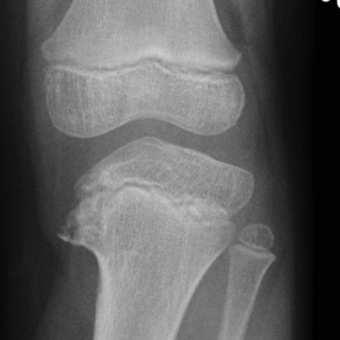

Figure 1

Figure 3


Radiological Analysis Report
1. Radiological Findings
Based on the provided X-ray images (anteroposterior and lateral views of both knees), the following observations can be noted:
- An obvious deformity and proliferation are observed at the proximal medial metaphysis of the tibia, leading to genu varum ("O"-shaped legs).
- Marked increase in bone density and deformity of the medial metaphysis with a "bony outgrowth"-like protrusion on the medial side (akin to the “beak” described in literature).
- The articular surface alignment is relatively acceptable, and no significant soft tissue thickening or calcification is visible.
- The bilateral lesions are largely symmetrical, with similar changes in the medial tibial aspects of both the right and the left sides.
Overall radiological manifestations are consistent with a medial proximal tibial lesion causing genu varum, suggesting the possibility of Blount's disease (tibial osteochondrosis).
2. Potential Diagnoses
Taking into account the patient’s gender (female), age (6 years), and clinical symptoms (bilateral mild knee pain and genu varum deformity), the following conditions are considered:
- Blount’s Disease (Tibial Osteochondrosis)
Typically presents with an abnormal medial proximal tibial metaphysis leading to genu varum, often appearing after the child begins walking and progressively worsening. X-rays may show a “beak”-like medial metaphyseal prominence and medial proximal tibial underdevelopment. - Physiological Genu Varum
Preschool-aged children may exhibit physiological genu varum; however, this usually corrects after two years of age. If there is no improvement or if it worsens after age 2–3, Blount’s disease should be ruled out. - Congenital Tibial Bowing
Usually presents with bowing at the mid or distal tibia and is often associated with other congenital anomalies, which is not consistent with the pronounced changes at the proximal medial epiphysis in this case. - Rickets
May cause skeletal deformities, typically showing cupping or fraying at the metaphyses or signs of osteopenia on radiographs, and is often accompanied by other manifestations of vitamin D deficiency. - Ollier Disease
Characterized by enchondromas; radiographically, multiple radiolucent areas within the bones can be seen, which do not align with the features in this case. - Others (e.g., traumatic or infectious growth plate injury, chondrodysplasia, etc.)
Require correlation with a history of trauma or local infection and other clinical evidence for exclusion.
3. Final Diagnosis
Considering the patient’s age, clinical presentation (genu varum, mild knee pain), possible epidemiological factors (higher prevalence in certain ethnic groups, early walking, family history), and the X-ray findings of medial metaphyseal deformity and protrusion, the most likely diagnosis is Blount’s disease (proximal tibial osteochondrosis).
4. Treatment Plan and Rehabilitation
For a child with confirmed or highly suspected Blount’s disease, early intervention is recommended to halt or slow the progression of the deformity:
- Conservative treatment:
- For patients in the early stage or with mild-to-moderate deformities, braces or orthoses can be used to help reduce excessive loading on the medial proximal tibia.
- Correct abnormal gait and posture; avoid premature or excessive weight-bearing.
- Surgical treatment:
- For severe or progressive deformities, tibial osteotomy or growth modulation surgery (e.g., an eight-plate procedure) may be considered to correct varus deformity and restore proper limb alignment.
- Postoperative management may include bracing, and rehabilitation should proceed gradually once bone healing is stable.
Rehabilitation and Exercise Prescription Recommendations:
- During the brace-wearing or postoperative recovery phase, start with basic range-of-motion (ROM) exercises and light muscle-strengthening to prevent joint stiffness or muscle atrophy from prolonged immobilization.
- In the initial stage, low or non–weight-bearing exercises can be performed, such as:
- Straight leg raises (supine or prone position) to strengthen the iliopsoas and quadriceps.
- Seated knee flexion and extension with low resistance, such as using a resistance band.
- Water-based training (swimming, ankle pump exercises at the pool’s edge), utilizing buoyancy to reduce stress on the knee joints, under professional guidance.
- Progressively increase weight-bearing and strength training:
- Gradually incorporate standing balance exercises under the supervision of a physical therapist, increasing standing time step by step.
- Lower-extremity resistance training (e.g., squats, step exercises) should be tailored to knee stability within a pain-free range, progressively increasing intensity and frequency.
- Adhere to the FITT-VP principle (Frequency, Intensity, Time, Type, Progression, Individualization):
- Frequency: 3–5 times per week.
- Intensity: Maintain low to moderate intensity as tolerated, avoiding high-impact loads.
- Time: Each session can last around 20–30 minutes, possibly divided into segments.
- Type: Water-based exercises, low-impact aerobic activities, or targeted functional training focusing on lower extremity strength and balance.
- Progression: Evaluate every 2–4 weeks and adjust accordingly. Once muscle strength and stability improve, gradually increase weight-bearing and activity level.
It is emphasized that patient safety is paramount during pediatric rehabilitation. Should any significant pain or discomfort arise, stop the activity immediately and seek medical evaluation.
Disclaimer: This report is based solely on the information currently provided and serves only as an initial reference. The final diagnosis and treatment should be determined by a clinical physician after a comprehensive assessment of the patient’s actual condition. It is not a substitute for in-person consultation or professional medical advice.
Human Doctor Final Diagnosis
Blount's Disease