Glomus tumor of the subungual region – MR and histological findings


Clinical History
A 41 year old patient presented with an enlarging painful mass at the subungual region of the right hallux. MRI exam was performed and the findings are presented.
Imaging Findings
A 41 year old female patient with a recent history of enlarging painful mass at the subungual region of the right hallux was referred to the radiology department by his GP. She was afebrile and no history of trauma in the region was referred. There was no history of diabetes. MRI exam was performed and demonstrated a mass located in the subungual region of the right hallux. On T1 weighted images (Fig. 1) the mass was hipointense to the muscular tissues. After admission of contrast medium the tumor was strongly enhancing (Fig. 2). On T2 fat-suppressed – weighted images (Fig ures 3, 4) the mass revealed an intense high MR signal. The patient underwent surgical excision of the tumor and the cytological examination (Fig. 5) revealed glomus tumor.
Discussion
Glomus tumors represent 1-5% of soft-tissue tumors in the extremities. In pathologic examination, glomus tumors are hamartomas developed from the neuromyoarterial glomus bodies that regulate blood flow in the skin.
The normal glomus body is an arteriovenous shunt that has an important role in thermoregulation and is present throughout the body in the deepest layer of the dermis, the reticular dermis. Each glomus body is an encapsulated oval organ of 300μm in length. It is located in the subungual region, digits and palms. Glomus bodies are highly concentrated in tips of the digits, particulary beneath the nails. The nail beds of fingers and toes contain 93–501 glomus bodies per square centimeter.
At clinical examination, glomus tumors are in majority non palpable lesions, frequently found in women. They usually provoke a compression to the nail matrix and thus, the principal symptom is pain at the region. A “classic triad” of clinical findings was described which include pain, point tenderness and cold sensitivity. The disappearance of the pain after application of a tourniquet proximally on the arm (Hildreth sign) is pathognominic of a glomus tumor. Eliciting pain by applying precise pressure with the tip of a pencil (Love test) helps locate the lesion.
Most glomus tumors are iso- or slightly hyperintense to the dermal layers of the nail bed on T1-weighted MR images and strongly hyperintense on T2-weighted images. The majority of them are surrounded by a capsule which has the appearance of a dark rim on T2-weighted and contrast material-enhanced imaging. The capsule is the result of a secondary reaction of the surrounding tissue and may be incomplete.
In some cases the signal intensity on T1WI and T2WI varies. It can be low or intermediate intense reflecting the variety of its histological components. The predominant cellular pattern of glomus tumors can be divided into 3 main forms: vascular , myxoid and solid. The solid or cellular type has a slightly high signal intensity on T2WI and injection of contrast medium helps its detection. The vascular type has a very strong enhancement. When the form of the tumor is myxoid the T2 times are very long. This scheme is theoretical because most tumors are composed of a mixture of the various types.
The differential diagnosis of glomus tumor includes mucoid cysts and angioma. Mucoid cysts are painless and at MRI imaging they present communication with the distal interphalangeal joint, longer values of T2 times and lack of contrast enhancement. The angioma may strongly mimic glomus tumor as they present the same signal intensity. Nevertheless, it is more superficial and located in the papillary dermis and the epidermis.
In our case, the tumor was located in the subungual region of the right hallux. Imaging techniques demonstrated the location of the tumor and its relation with the subsequent tissues. The cytological examination revealed the nature of the tumor and its composition of vascular, myxoid and solid components.
Differential Diagnosis List
Final Diagnosis
Glomus tumor of the subungual region of the right hallux
Liscense
Figures
T1WI, coronal plan

T1WI, coronal plan, after admission of contrast medium
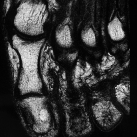
T2 fat-suppressed weighted image, axial plan

T2 fat-suppressed weighted image, saggital plan

Histologic speciment

1. Imaging Findings
The patient is a 41-year-old female, presenting with a gradually enlarging painful mass located under the nail (subungual region) of the right big toe. Based on the provided MRI images, the following key features are noted:
- Lesion location: The lesion is situated in the subungual region of the right big toe, appearing as a focal occupying lesion.
- Morphology and signal: On T1-weighted imaging, the lesion shows signal intensity similar to or slightly higher than the surrounding nail bed tissue; on T2-weighted imaging, it appears markedly hyperintense, consistent with vascular-rich or fluid-rich tissue.
- Pseudocapsule sign: In some sequences, a low-signal rim (resembling a thin “capsule”) can be seen around the periphery of the lesion, suggesting a reactive boundary formed by surrounding tissues, although this capsule may be incomplete.
- Enhancement characteristics: According to the literature and typical presentations, such lesions often exhibit prominent enhancement after contrast administration, especially in areas with abundant vascularization.
- Surrounding structures: The lesion is located beneath the nail bed, exerting some degree of pressure on the nail matrix and adjacent soft tissue, but no significant bone destruction is observed.
2. Potential Diagnoses
Based on the patient’s history (no prior trauma, female, painful subungual mass) and the MRI findings (signal characteristics), the main differential diagnoses include:
- Glomus tumor: Commonly found in the subungual region, often accompanied by severe pain and sensitivity to cold. On MRI, it may appear isointense or mildly hyperintense on T1, hyperintense on T2, and shows pronounced enhancement. This aligns well with the patient's symptoms and imaging features.
- Mucoid cyst: Typically connected to the distal interphalangeal joint. It shows higher T2 signal, often does not present with severe pain, and may not show significant enhancement. Clinically, it usually presents with milder pain.
- Hemangioma: May demonstrate a similarly high signal on imaging but is more commonly encountered in superficial skin layers (dermis and epidermis) rather than deep within the subungual space, and clinical features differ accordingly.
3. Final Diagnosis
Taking into account the patient’s age, clinical presentation (painful subungual mass, possible cold sensitivity), MRI features (signal and enhancement patterns), and the pathological examination revealing abundant vascular components as well as mucoid and solid elements, the most consistent diagnosis is:
Glomus tumor
4. Treatment Plan and Rehabilitation
Treatment Strategy:
- Surgical excision: For symptomatic glomus tumors, surgery is the primary and effective treatment. Complete removal following accurate lesion localization can significantly alleviate pain and reduce the risk of recurrence.
- Postoperative care: Proper wound care is essential, as is avoiding excessive pressure or trauma to the area to promote healing.
Rehabilitation and Exercise Prescription:
- Early phase (1–2 weeks post-surgery):
- Focus on wound care and gentle toe movements, minimizing external pressure or impact.
- If walking is necessary, wear loose-fitting footwear or use supportive devices to reduce local stress.
- Mid phase (2–4 weeks post-surgery):
- Gradually increase the range of motion and active toe movements, such as seated flexion and extension exercises.
- Recommended frequency and duration: 2–3 times a day, 5–10 minutes each time, adjusted based on pain tolerance.
- If there is no significant pain or swelling, moderate walking can be introduced; however, avoid strenuous running or jumping.
- Late phase (4 weeks post-surgery and onwards):
- Depending on the healing status, progressively introduce weight-bearing activities like slow-paced walking or low-impact aerobics.
- For strengthening, use resistance band exercises or balance training for the toes under professional guidance.
- Follow the FITT-VP principle (Frequency, Intensity, Time, Type, Volume, Progression), adjusting according to individual tolerance.
- Individual Considerations:
- Seek prompt medical attention if pain, swelling, or bleeding persists or worsens.
- Patients with chronic health issues such as diabetes or peripheral vascular disease should coordinate with relevant specialists to ensure optimal wound healing.
Disclaimer
This report serves as a reference analysis, based on currently available imaging and pathological data, as well as related medical literature. It does not replace in-person consultation or professional medical advice. Should you have any questions or changes in your condition, please seek immediate medical attention or consult specialized healthcare providers for further examination and treatment.
Human Doctor Final Diagnosis
Glomus tumor of the subungual region of the right hallux