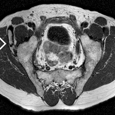
Pelvic finger: X-Ray and MR imaging


Clinical History
Vague chronic pain referred to lumbar spine and hips.
Imaging Findings
The patient consulted for chronic lumbar and pelvic pain. Neither pelvic surgery nor trauma were recorded in the past medical history. His physical examination was anodyne. Laboratory tests were normal. Plain radiographs of lumbar spine and both hips were obtained. Anterior posterior and oblique films of the right hip showed a bony structure attached to the anterior inferior iliac spine, with two segments. Pelvic MR was performed to confirm the findings of X-ray films, the presence of bone marrow, and the pseudo-joints between the two bones and with the anterior inferior iliac spine.
Discussion
Pelvic digit, also named pelvic rib or iliac rib, is an extremely rare development anomaly. Bone structures develop in the soft tissues close to the osseous pelvis (ischium, ilium, sacrum or coccix). Sometimes it has a rib-like appearance, but the characteristic is a phalanx-like structure with one or more pseudo-joints within and at the base. “Iliac digits” seem to be more frequent, in particular at the anterior inferior iliac spine, as in this case. The pelvic digit is usually asymptomatic and there is absence of history of trauma. Sometimes occurs bilaterally. This anomalous structure must arise from the embryonic mesoderm with rib-forming capacity present in these areas, that failures to involute. Plain radiograph appearance is typical. In equivocal cases, cross-sectional imaging, such as Magnetic Resonance, can be performed to confirm the presence of cortical bone, bone marrow, and the pseudo-joints. Differential diagnosis include myositis ossificans, avulsion injuries, osteochondroma or Fong's disease (bilateral iliac horns). In conclusion, pelvic digit is an asymptomatic benign condition. It must be kept in mind when an atypical bone structure is noted close to the pelvis, and therefore avoid additional investigations.
Differential Diagnosis List
Final Diagnosis
Pelvic digit
Liscense
Figures
Anterior posterior plain radiograph of the right hip

Oblique plain radiograph of the right hip

Paraaxial T1-weigthed scan of the pelvis

Sagital T1-weighted scan of the right hip

Fat-supressed T1-weighted axial of the right pelvis

Imaging Report
I. Imaging Findings
From the provided pelvic X-ray and MRI images, it can be observed that in the soft tissue near the right (or possibly bilateral, depending on careful scrutiny of the actual images) iliac bone, there is one or multiple abnormal bony structures shaped like “finger-like” or “rib-like.” These bony structures demonstrate:
- Signals indicating both cortical bone and bone marrow components;
- A connection to normal bone or a pseudo-joint-like interface on the surface;
- No obvious soft tissue swelling or inflammatory signal changes, and no erosive or destructive features.
In addition, this patient shows no clear evidence of fracture or other notable bony destruction. There are no prominent soft tissue masses or signs of acute local injury seen on the imaging studies.
II. Possible Diagnoses
Based on the patient's age, symptoms (chronic mild pain), and imaging characteristics, the following differential diagnoses can be considered:
- 1. Pelvic Digit / Congenital Bony Protuberance near the Ilium:
Also referred to as an “iliac rib” or “pelvic rib.” Commonly appears as a finger-like or rib-like protrusion, potentially forming a pseudo-joint around the pelvic region. It is a rare congenital developmental anomaly, usually asymptomatic. - 2. Myositis Ossificans:
Often occurs after trauma or inflammation leading to ossification within soft tissue. Radiologically, it usually manifests as irregular calcification within the soft tissue, typically with a history of trauma or other precipitating factors, and the calcification often has a concentric zonal pattern. - 3. Osteophyte / Osteochondroma:
Commonly seen as an outward growth from the bone surface, often with a cartilage cap, usually near the growth plate. It typically appears “mushroom-like” or “club-like.” - 4. Avulsion Injury:
Most frequently seen in adolescents or athletes. Sudden pulling forces or trauma at muscle attachment sites on the pelvis can result in avulsion fractures. Imaging often shows fracture lines and irregular bone cortices, typically accompanied by acute pain. - 5. Fong Disease (Bilateral Iliac Horns):
Can be associated with the nail-patella syndrome; usually presents as bilateral “horn-like” projections of the iliac bones.
III. Final Diagnosis
Considering the patient’s long-standing mild chronic pain without a typical history of trauma or inflammation, combined with imaging findings of a “finger-like” bony structure and pseudo-joint-like changes, the findings strongly suggest:
“Pelvic Digit,” a congenital benign developmental anomaly.
This condition is typically asymptomatic and is often discovered incidentally. Further confirmation can be achieved through high-resolution CT or close clinical observation, but the current data are sufficient to support this diagnosis.
IV. Treatment Plan and Rehabilitation
In most cases, a pelvic digit is a benign congenital variation. If there are no significant symptoms or complications, no special treatment is required. If the patient experiences localized pain or discomfort, the following measures may be considered:
- Conservative Treatment:
- Oral or topical non-steroidal anti-inflammatory drugs (NSAIDs) to alleviate chronic local pain symptoms.
- Physical therapy and rehabilitation exercises: including local heat therapy, massage, and stretching to reduce muscle tension.
- Lifestyle adjustments: avoid prolonged unilateral weight-bearing or maintaining fixed positions, and schedule regular movements to prevent muscle fatigue.
- Surgical Treatment:
Considering surgical resection only if this structure repeatedly causes irritation or, in very rare circumstances, impacts function. Such cases are extremely uncommon.
For general low back and pelvic area chronic pain, a gradual exercise regimen can help improve soft tissue endurance and muscle strength, reducing compensatory pain.
- F (Frequency): 3–4 sessions of exercise per week.
- I (Intensity): begin with low to moderate intensity (subjective effort level 4–5 out of 10), avoiding sudden heavy loads.
- T (Time): each exercise session should last approximately 20–30 minutes initially and can be gradually extended up to 45 minutes as tolerated.
- T (Type): primarily focus on core muscle strengthening and back muscle exercises, supplemented by low-impact aerobic exercises such as brisk walking, swimming, or using an elliptical machine.
- V (Volume): combine the number of weekly sessions with the duration of each session, keeping it within a reasonable range without significant increases in a short period.
- P (Progression): progressively increase load or duration as guided by perceived pain and fatigue levels, and incorporate deeper core training such as planks or bridge exercises as appropriate.
If the patient also has osteoporosis or other underlying conditions, exercise should be conducted under the guidance of a professional physician or physical therapist, with close monitoring of any discomfort or pain during and after workouts.
Disclaimer
This report is provided for reference only and cannot substitute for individual assessment or the diagnostic opinion of a professional physician in a clinical setting. If you have further questions or experience worsening symptoms, please consult a specialist promptly.
Human Doctor Final Diagnosis
Pelvic digit