Aplasia of the posterior arch of the atlas with a remnant tubercle and a cleft of the anterior arch.


Clinical History
Cycling athlete, referred head injury.
Imaging Findings
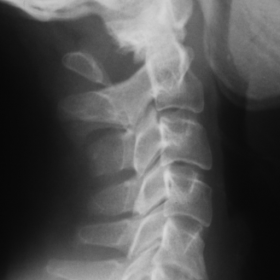
A 31 year old Caucasian male - a cycling athlete - was admitted to the hospital after neck and head injury. Physical examination of the cervical spine, as well as neurologic examination, was normal. The lateral radiograph revealed an interrupted posterior arch of the atlas and presence of a posterior remnant (Fig 1). This was initially misinterpreted as a fracture, but a more detailed inspection showed that the defect had sclerotic borders. CT revealed the absence of ossification of both posterior arches of the atlas, the presence of an intact posterior tubercle and also demonstrated a cleft in the anterior arch (Fig 2,3). There was no soft tissue involvement or bony injury.
Discussion
Atlas vertebra consists of an anterior and a posterior arch, each of which has a tubercle and a lateral mass. During embryonic period, atlas develops from three ossification centres, one mid-line anterior centre which forms the anterior arch and two lateral centres for each lateral mass [1,2]. The ossification centre for the anterior arch and tubercle is unified with the lateral masses at 5-9 years of age [2,3]. The posterior arch is created by perichondral growth unification of two lateral masses centres by 3-5years [1,2,3]. However, in 2% of the population a 4th centre forms the posterior tubercle [4]. Congenital anterior defect cleft vertebra is due to variation or absence of the anterior ossification centre and failure for the lateral masses to fuse anteriorly [5]. Congenital posterior arch abnormalities range from small clefts to complete absence. Developmental deficiencies of the posterior arch imply a defective or absent cartilaginous preformation and not a disturbance of the ossification [1,2,6]. Consequently, bony deficits are bridged by loose connective tissue instead of cartilage [6]. Most atlas anomalies produce no abnormal craniovertebral junction relationships and are not associated with platybasia. The reported incidences are 4% for posterior and 1% for anterior arch defects [2,6]. Abnormalities associated with absence of the posterior arch may be fusion of the vertebra C2-C4 (Klippel-Feil syndrome) and prominence of the posterior tubercle of the axis, bilateral clefts of the axis and an elongated articular process of C3 [2]. Hypoplastic posterior arch of the atlas may be seen in children with Down syndrome, in association with atlantoaxial dislocation, amplifying the risk of spinal cord damage.
According to Currarino et al [1], posterior arch defects are divided into five groups: Type A results from failure of the posterior mid-line fusion of the two hemiarches and accounts more than 90% of all posterior arch defects; type B includes unilateral clefts, ranging from a small cleft to the complete absence of the hemi-arch; type C corresponds to bilateral clefts; type D, as in our case, includes absence of the posterior arch associated with a persistent posterior tubercle. Finally, type E refers to absence of the entire posterior arch including the posterior tubercle, often associated with hypertrophy of the spinous process of C2. In most cases these defects are disclosed incidentally in patients having cervical spine radiographs, usually after trauma. The main clinical implication includes potential misdiagnosis as fractures [3]. Quadriparesis following minor cervical or head trauma and Lehrmitte sign may be seen in a minority of patients with posterior arch aplasia and it is attributed to an impingement of the tubercle on the spinal cord during extension [3,4,6]. In the presence of neurological symptoms, MRI may reveal cord contusion [3].
The congenital clefts, as opposed to fractures, have smooth and sclerotic borders on radiographs, without soft tissue swelling. CT imaging with a 3D reconstruction is extremely helpful in demonstrating these spinal anomalies.
Differential Diagnosis List
Final Diagnosis
Bilateral atlas posterior arch aplasia, remnant tubercle, anterior arch cleft.
Liscense
Figures
Lateral radiograph of the cervical spine

Axial CT of the atlas

3-D reconstruction of the atlas

1. Imaging Findings
Based on the lateral cervical X-ray, axial CT, and three-dimensional reconstruction images, it is evident that the posterior bony structure of the first cervical vertebra (atlas) is missing, with the posterior arch not fully visualized. Part of the posterior tubercle is present but lacks continuity with the posterior arch. CT 3D reconstruction images clearly demonstrate a congenital defect in the posterior portion of the atlas, with smooth sclerotic edges and no signs of soft tissue swelling or fracture lines, suggesting a congenital deficiency rather than a traumatic fracture.
2. Possible Diagnoses
Considering the patient’s age (31 years), medical history (head injury while cycling), and imaging features, the following should be differentiated:
- Congenital Posterior Arch Defect of the Atlas (Currarino Classification Type D): Incomplete or absent posterior arch with a residual posterior tubercle. Margins are smooth, and there are no signs of fracture.
- Traumatic Fracture of the Atlas: A fracture would usually show irregular fracture lines and surrounding soft tissue swelling; patients often present with severe pain or neurological symptoms. No clear evidence of trauma is indicated in this case.
- Other Congenital Variations of the Cervical Spine: Such as C2 vertebral or lower cervical fusions. However, current images primarily show a defect in the posterior arch of the atlas, with no evident fusion abnormalities in other vertebrae.
Given the imaging findings of smooth margins and no cortical disruption, along with literature suggesting congenital posterior arch defects of the atlas are not uncommon (approximately 4%), a congenital posterior arch defect is highly suspected.
3. Final Diagnosis
Combining the patient’s clinical presentation, imaging findings, and related literature, the most likely diagnosis in this case is:
Congenital Posterior Arch Defect of the Atlas (Currarino Classification Type D).
If further evaluation of cervical spine stability or potential nerve compression is required, additional dynamic cervical X-rays or MRI should be considered.
4. Treatment and Rehabilitation Plan
For patients with a congenital posterior arch defect of the atlas and no neurological deficits or evident cervical instability, conservative treatment and routine follow-up are generally recommended, including:
- Conservative Observation: If asymptomatic or presenting only mild neck discomfort, no special interventions are typically needed. Periodic imaging follow-ups can be used to monitor cervical stability.
- Temporary Neck Brace: In the early phase of rehabilitation after the recent trauma, a short-term use of a cervical collar may be considered to limit cervical motion and reduce the risk of further injury.
- Surgical Indications: If significant neurological symptoms (e.g., limb weakness, spinal cord compression) develop or imaging suggests cervical instability, surgical fusion or decompression should be considered.
Rehabilitation/Exercise Prescription Recommendations: In the absence of significant neurological symptoms or bony instability, mild cervical functional rehabilitation exercises can be undertaken. Refer to the FITT-VP principle for guidance:
- Frequency: 3–4 times a week initially, gradually increasing to simple neck exercises daily.
- Intensity: Focus on low to moderate intensity, with slow movements and avoidance of sudden neck rotations or excessive extension.
- Time: Start with 10–15 minutes per session, gradually extending based on patient tolerance.
- Type: Include active movements such as flexion, extension, lateral bending, and rotation of the neck; consider adding shoulder girdle relaxation or low-resistance exercises as appropriate.
- Progression: Increase range of motion and resistance gradually as symptoms improve and tolerance increases. If well tolerated, core muscle and upper extremity strengthening exercises can be added to ensure stability of the neck-shoulder region.
Close attention should be paid to any increase in neck pain or signs of nerve compression (e.g., tingling, numbness, weakness) during rehabilitation. If such symptoms occur, seek medical advice promptly.
Disclaimer: The above report is a preliminary medical analysis based on limited information and is for reference only. It should not replace an in-person consultation or professional medical advice. If you have any concerns or worsening symptoms, please seek medical attention.
Human Doctor Final Diagnosis
Bilateral atlas posterior arch aplasia, remnant tubercle, anterior arch cleft.