Painful foot lump!


Clinical History
The patient was referred for a painful growing mass of the foot.
Imaging Findings
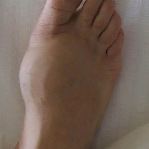
A 43 year-old man presented with a 6-month history of a painful growing mass on the dorsum of the right foot (Fig 1). An AP radiograph revealed an expansible, eccentric, well-defined bone lytic lesion, with barely sclerotic margins (Fig 2). MR (FSE sequences) showed an expansible nodular mass of the 1st metatarsal bone, abutting the articular surface, with marked low signal on T1-weighted images and higher and inhomogeneous signal on T2-weighted images (Fig 3). Bone scanning revealed no metastatic disease (Fig 4). Angiography reconfirmed its hypervascular nature (Fig 5). The differential diagnosis included foot enchondroma, giant cell tumour (CGT), aneurysmal bone cyst (ABC), chondroblastoma and intraosseous ganglion, Brodie abscess.Surgery was performed (Fig 6) and histologic examination revealed a bone giant cell tumour (Fig 7).
The patient recovered well after surgery.
Discussion
Giant cell tumour (GCT) is a common neoplasm (4-5% of primary bone tumours) and consists of connective tissue, osteoclastic giant cells and a fibrous stroma. It's one of the few bone tumours that has a slight predilection for females (1.5:1). Even though GCT can be clinically aggressive, neither its radiographic nor histologic appearance can put them into a benign or malignant category. After surgical excision, they are considered benign unless they recur (5-15% are thought to be malignant). Malignant GCT can metastize to the lungs, and less frequently, to lymph nodes. Despite the presence of metastatic disease, the prognosis remains good, with the major difficulty being that of local recurrence.
Patients may present with pain, swelling, limited range of motion of the adjacent joint or, with a pathologic fracture.
Typically, lesions occur in the long bones (distal femur, proximal tibia, distal radius and ulna or proximal humerus). Additional sites of involvement include proximal femur, sacrum, ribs, vertebrae, hands and feet (talus and calcaneus are the most commonly affected, followed by metatarsal bones).
There are 4 classic radiographic criteria for diagnosing GCT in long bones:
1) mainly affects skeletally mature patients, with 80% occurring in patients between 20 and 50 years.
2) even though the exact site of origin of GCT has been controversial, when the radiologist sees this lesion it's already epiphyseal and abuts the articular surface.
3) eccentrically located.
4) associated with a narrow zone of transition and lacking surrounding sclerosis (80%–85% of patients, Lodwick 1B).
These rules do not apply in flat bones or in the apophyses, which have no articular surfaces and can have a sclerotic margin (Lodwick 1A).
Other useful, frequent criteria are a slightly expansile nature and the not unusual extension of the tumour into the soft tissue. Also, the absence of periosteal reaction (unless a fracture has occurred) and the lack of mineralized matrix should be kept in mind. The age of the patient and the lack of mineralized matrix help to distinguish GCT from other end-of-bone lesions. Cystic (secondary ABC) components, frequently with fluid levels levels resulting from haemorrhage and necrosis are common (14% of lesions), along with solid portions (the presence of both solid and cystic components allows distinction from primary ABC, which contains only cystic regions).
CT improves detection of cortical thinning, pathologic fracture, periosteal reaction, lack of mineralization and degree of osseous expansile remodeling compared with radiography.
MRI is superior to CT in delineating soft-tissue tumor extent. It reveals a relatively well-defined lesion with a low-signal-intensity margins, with low to intermediate signal intensity at T1- and T2WI in the majority of cases and a heterogeneous pattern of enhancement after gadolinium. This feature can be useful in excluding other subarticular lesions such as solitary subchondral cyst, intraosseous ganglion, Brodie abscess, and clear cell chondrosarcoma that demonstrate high signal intensity at T2WI. Areas of very low signal intensity on all sequences are not unusual and are due to deposition of hemosiderin.
Differential Diagnosis List
Final Diagnosis
Bone giant cell tumour of the foot (first metatarsal bone)
Liscense
Figures
foot lump
AP radiographic view

MRI


Bone scanning

Surgical specimen

histologic examination

Digital Subtraction Angiography

1. Imaging Findings
The patient is a 46-year-old male complaining of a painful, progressively enlarging mass in the foot. Based on the provided X-ray, MRI, and other imaging studies, the following observations can be made:
- In the foot region (commonly in the tarsal or metatarsal areas), a distinct lytic lesion is visible, with relatively clear boundaries and slightly expansile changes.
- X-ray findings suggest the lesion is close to the articular surface and exhibits an eccentric distribution. The cortical bone may appear thinned without obvious sclerotic margin, and there may be limited soft tissue swelling.
- MRI reveals that the lesion shows low to intermediate signal on T1-weighted images and a mixed or intermediate signal on T2-weighted images. Septations or cystic components are noted, suggesting possible secondary aneurysmal bone cyst-like changes, while the lesion margins remain intact.
- No evident mineralized matrix or significant periosteal reaction is observed.
- Post-contrast enhancement shows marked enhancement in the solid portion of the lesion, while the cystic region shows no enhancement.
2. Potential Diagnoses
Taking into account the patient’s age, clinical presentation, and imaging characteristics, the following differential diagnoses could be considered:
- Giant Cell Tumor (GCT):
Typically occurs in individuals aged 20–50 years, often near the articular surface of long bones, but can also be found in tarsal or metatarsal bones. It presents as an aggressive lytic lesion with well-defined borders and usually no significant sclerosis. - Aneurysmal Bone Cyst (ABC):
Characterized by a multi-chamber expansile lytic lesion, often described as “blow-out” or “soap bubble” in appearance, with septations or fluid-fluid levels. Pure ABCs, however, are less likely to have extensive solid components and more commonly occur in patients with open growth plates, which does not fully align with this case’s age and location. - Chondroma or Chondrosarcoma (e.g., clear cell chondrosarcoma):
In cases of chondrosarcoma, X-ray images often show characteristic cartilaginous calcifications (ring-shaped or stippled). These are not observed in this case, making this diagnosis less likely. - Benign bone cyst or disseminated lesions:
A single, eccentric lesion lacking a surrounding sclerotic rim is not highly suggestive of a simple bone cyst or metastatic lesion.
3. Final Diagnosis
Considering the patient’s age, symptoms, and imaging features (a lytic lesion with relatively clear borders but slightly aggressive characteristics, proximity to the articular surface, mixed signals on MRI with solid enhancement, and lack of obvious calcification), along with references from existing literature and pathological examination, the most probable diagnosis is:
Giant cell tumor of the foot (Giant cell tumor of bone).
If uncertainty remains, a biopsy or intraoperative frozen section can be performed to further clarify the nature of the lesion.
4. Treatment Plan and Rehabilitation Program
Treatment Strategy:
- For early or well-localized lesions, surgical intervention may be considered, such as curettage of the lesion, or curettage combined with bone grafting/internal fixation. In some cases, extensive resection or reconstructive surgery may be required.
- If there is soft tissue involvement, a large lesion, or uncertain margins, preoperative embolization or adjuvant radiotherapy could be considered before surgery. Chemotherapy typically shows limited efficacy for classic giant cell tumors; however, it can be considered for malignant or metastatic forms after assessment by a multidisciplinary team (MDT).
- Monoclonal antibodies against osteoclasts (e.g., Denosumab) may be used in treating giant cell tumors of bone, especially in cases not suitable for surgery or as a postoperative measure to prevent recurrence.
Rehabilitation and Exercise Prescription (FITT-VP):
- Frequency (F): In the early recovery phase after treatment, perform light rehabilitation exercises 1–2 times a day to promote local blood circulation and soft tissue healing.
- Intensity (I): Start with non-weight-bearing or partial weight-bearing exercises, such as foot stretching and ankle range-of-motion (ROM) training, avoiding high impact.
- Time (T): Each training session should last 10–15 minutes initially, gradually increasing to 20–30 minutes as recovery progresses.
- Type (T): Options include swimming, lower limb strengthening (e.g., wall sits with limited foot load), joint mobilization exercises, and low-impact aerobic activities (e.g., stationary cycling).
- Progression (P): As the lesion heals and pain improves, progressively increase weight-bearing and intensity. A reassessment every 1–2 weeks is recommended to steadily advance activity levels.
- Volume and Physical Fitness (V-P): Gradually increase the overall training volume, avoiding excessive fatigue or pain exacerbation. Regularly monitor bone healing and soft tissue status, and use additional bracing if necessary.
Given that giant cell tumors have a certain recurrence rate, periodic imaging follow-up is essential during rehabilitation to closely observe bone and soft tissue healing. Adjust the intensity of training as needed.
Disclaimer: This report is a reference-based analysis using existing information and cannot replace an in-person consultation or professional medical advice. Specific treatment and rehabilitation plans should be formulated by a professional medical team based on the individual patient’s condition.
Human Doctor Final Diagnosis
Bone giant cell tumour of the foot (first metatarsal bone)