Bilateral Madelung\'s deformity


Clinical History
A 25 year old lady started complaining of increasing pain and deformity of both wrist over a few months. No history of trauma was evident. She was referred to the orthopaedic outpatient clininc by her GP.
Imaging Findings
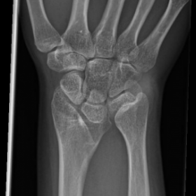
25-year-old lady presented to the outpatient clinic complaining of increasing pain and deformity of both wrists. Inspection revealed the prominence of the distal ulna and bowing of the radius on both sides. Examination revealed the presence of marked tenderness over the distal radioulnar and wrist joints. Both active and passive joint movements were limited on both sides. Physical examination revealed short stature. Standard AP and Lateral plain X-ray projections of both forearms were taken.
Discussion
This condition known as carpus curves results from epiphyseal growth arrest on the volar and ulnar half of the distal radius, causing the articular surface to be directed ulnar and volarward. It is inherited as an autosomal dominant condition with variable expressivity. It shows a predominantly female distribution with a 4:1 ratio. It is bilateral in two thirds of patients.
It is caused by the complete absence or underdevelopment of the ulnar portion of the radius growth plate, thus failing to contribute to the linear growth of the corresponding border of the radial diaphysis. The precise mechanism of this disorder has not been determined however hemiarthropathy of the distal radius epiphysis, abnormal muscular insertions and disturbances to the vascular supply of the epiphysis have all been suggested.
It is associated with Madelung’s dyschondrosteosis and mesomelic dwarfism should be suspected if the condition is bilateral.
Clinically it is characterised by insidious onset of pain in one wrist, then in other, and increasing prominence of the dorsal ulnar head and bowing of distal radius. Pain from radioulnar subluxation or radiolunate impingement usually becomes less severe as the patient grows older. However when the deformity becomes stabilised, the incongruity of the joint surfaces in the wrist may lead to the recurrence of painful symptoms. Deformity progresses until growth plate of the distal radius closes. Wrist motion, particularly extension and supination, is limited. Although Madelung's deformity is considered a congenital deformity, it does not become manifest until in late childhood or early adolescence.
Treatment options include non operative conservative measures indicated early on in the condition especially when the patient is asymptomatic. Various surgical procedures are available in the treatment plan for this condition.
Differential Diagnosis List
Final Diagnosis
Madelung's deformity
Liscense
Figures
RT AP

RT LAT

LT AP

LT LAT

Medical Imaging Analysis Report
I. Imaging Findings
Based on the provided bilateral wrist X-ray images, there is noticeable deformity of the bilateral distal radius. The distal articular surface tilts toward the ulnar and palmar side (i.e., the closure line of the distal radius shifts toward the ulnar and palmar sides). Additionally, there seems to be some asymmetry in the distal radioulnar joint surface, with the dorsal prominence of the ulnar head. The radial shaft may exhibit a certain degree of curvature, leading to an imbalance in the length proportion between the radius and ulna, making the ulna relatively more prominent. These findings are consistent with typical features of Madelung’s deformity (also known as “distal radial growth plate abnormality”).
II. Possible Diagnoses
- 1. Madelung’s deformity: This condition commonly presents as impaired growth of the distal radius, causing tilt of the distal radial articular surface to the ulnar and palmar sides, often accompanied by relative prominence of the ulnar head. It is more frequent in young females and can be bilateral. This aligns with the patient’s age, gender, and imaging findings.
- 2. Other congenital or genetic skeletal developmental abnormalities: Conditions such as partial skeletal dysplasia or chondrodysplasia can present similarly, though they generally involve other skeletal abnormalities. Based on clinical correlation, Madelung’s deformity is more likely.
- 3. Post-traumatic premature physeal closure or malunion of the distal radius: If there were a clear history of trauma, secondary deformity could arise due to malunion. Since no trauma history is reported, this possibility is less likely.
III. Final Diagnosis
Considering the patient’s profile as a 25-year-old female, the gradual onset of bilateral wrist pain and deformity, typical distal radial tilt, and dorsal prominence of the ulna, the most likely diagnosis is:
Madelung’s deformity.
For further clarification of the degree of deformity and evaluation of soft tissue, CT reconstruction and three-dimensional assessment, or MRI for cartilage and ligament evaluation, may be considered if necessary.
IV. Treatment Plan and Rehabilitation
Treatment strategy depends on the severity of the symptoms, progress of the deformity, and extent of functional impairment. Generally, it can be divided into conservative management and surgical intervention:
- Conservative Management: Suitable for patients with mild deformity or insignificant symptoms. Possible measures include:
- Wearing splints or wrist supports: To stabilize the wrist, reduce pain, and prevent progression of deformity.
- Anti-inflammatory and analgesic medications: For acute phases or significant pain, taken orally or applied topically for a short duration.
- Physical therapy: Such as heat therapy, ultrasound, iontophoresis, to help relieve pain and promote local blood circulation.
- Surgical Intervention: Recommended for severe deformities, significantly impaired wrist function, or persistent pain and instability. Surgical procedures may include radial osteotomy, ulnar shortening, or joint reconstruction, depending on the patient’s specific condition.
Rehabilitation/Exercise Prescription Recommendations (FITT-VP Principle):
For patients with wrist dysfunction and associated pain, rehabilitation focuses on gradually restoring joint range of motion, strengthening the muscles around the wrist, and enhancing stability:
- Frequency: It is recommended to perform wrist rehabilitation exercises 3–5 times per week.
- Intensity: Start with low intensity exercises (e.g., light resistance training using small resistance bands), progressing gradually according to pain and swelling.
- Time: Begin with 10–20 minutes of training per session, potentially split into segments. As tolerance improves, gradually increase to 30 minutes.
- Type:
- Range of Motion Exercises: Such as active flexion, extension, pronation, and supination, combined with gentle stretching.
- Muscle Strengthening: Use light dumbbells or resistance bands for wrist extension, flexion, and forearm rotation exercises. Avoid excessive range or force.
- Proprioception and Stability Training: For instance, using grip balls or stability balls under safe conditions.
- Progression: Adjust resistance and repetition every 2–4 weeks based on exercise tolerance, avoiding sudden excessive loads that may lead to reinjury.
- Volume & Personalization: Customize according to the patient’s level of wrist pain, severity of deformity, and muscle strength of supporting structures.
Disclaimer
This report is based solely on the provided imaging and patient history for preliminary analysis and does not replace a face-to-face clinical diagnosis or expert medical guidance. If you have further questions or if symptoms worsen, please seek prompt medical attention and follow a treatment and rehabilitation plan under the guidance of a specialist.
Human Doctor Final Diagnosis
Madelung's deformity