Chronic Recurrent Multifocal Osteomyelitis (ECR 2010 Case of the Day)


Clinical History
An 18-year-old female presented with pain in the left clavicle. She was known with intermittent pain and swelling on the medial clavicle and right ribs in the last four years, nonresponsive to local corticoid. The histology and culture of two previous percutaneous biopsies were inconclusive. The laboratory tests were unremarkable.
Imaging Findings
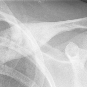
Radiographs (anteroposterior clavicle and posteroanterior chest) revealed predominantly sclerotic lesions with a minor lytic component, and marked expansion and solid periosteal reaction on the medial two-thirds of the left clavicle and anterior aspect of the 6th right rib (Fig. 1-3).
Tc-99m methylene diphosphonate bone scintigraphy (anterior projection) demonstrated increased uptake in the locations previously described as well as in the 5th right rib, the latter being radiographically occult (Fig. 4).
Magnetic resonance imaging (MRI) of the left clavicle depicted a medullary lesion with sclerosis and multiple patchy and coalescent areas isointense in T1-weighted images (WI), hyperintense in T2-WI, and strongly enhancing after gadolinium on the medial two-thirds of the clavicle. The solid periosteal reaction is also appreciated as well as soft tissue edematous changes (Fig. 5-8).
Discussion
Chronic recurrent multifocal osteomyelitis (CRMO) is a rare, autoinflammatory disorder typically affecting children and adolescents. CRMO belongs to the spectrum of aseptic osteomyelitis-like conditions in which SAPHO (Synovitis, Acne, Pustulosis, Hyperostosis, and Osteitis) syndrome is included. Its cause remains unknown though recently a genetic component has been suggested. Its incidence is estimated to be 2–5% of all osteomyelitis cases [1, 2]. CRMO is twice as common in females and the age of onset is usually 4–14 years although it has been reported in adulthood [1, 2, 3]. The main complaints are localized pain and variable swelling, possibly associated with mild malaise and dermatologic affections as palmoplantar pustulosis [2, 3].
Lesions are typically bilateral and multifocal at presentation and involve the metaphyses of tubular bones and medial clavicle, and also commonly the spine, ribs, mandible and pelvis [1, 2, 4]. CRMO has a prolonged course with relapses and remissions, involving the same or a different location. Histopathology is that of a non-specific inflammation, with fibrosis and/or hyperostotic regeneration [5].
The mainstays of imaging diagnosis are radiography and bone scintigraphy. Initially, the lesions are purely osteolytic developing variable sclerosis and periosteal reaction over time. After clinical resolution, radiographic appearance can normalize in 0,5-5 years or maintain remnants of the bony alteration. Complete healing of the clavicular lesions is rare [2, 3, 4]. MRI is highly sensitive for diagnosing occult lesions and whole body MRI is increasingly used for evaluation of multifocal bone lesions. MRI is extremely accurate to evaluate disease activity and local extent, being helpful for biopsy guidance and evaluation of long-standing disease. Active disease manifests as areas of high signal on T2-WI and enhancement. Quiescent disease correlates with low signal on T1- and T2-WI corresponding to sclerosis, possibly with scattered areas of high signal related to fatty marrow [1, 2].
Owing to the lack of specific tests the diagnosis of CRMO is one of exclusion. Although CRMO can be self-limited, it can have a protracted course and result in significant morbidity. The multifocal involvement of typical sites particularly the anterior thoracic wall, as in the case in appreciation, is crucial to accurate diagnosis.
Radiologists can be the first to suggest CRMO diagnosis given its characteristic radiographic appearance and distribution, thus preventing unnecessary multiple biopsies and long-term antibiotic treatment.
Differential Diagnosis List
Final Diagnosis
Chronic Recurrent Multifocal Osteomyelitis
Liscense
Figures
anteroposterior clavicle radiograph

posteroanterior chest radiograph

rib detail

Tc-99m methylene diphosphonate bone scintigraphy

MRI of the left clavicle. Axial T1W image

Axial T2W fat sat image

Axial T1W fat sat after i.v. contrast

Sagittal T1W fat sat after i.v. contrast

Medical Imaging Analysis Report
I. Imaging Findings
The patient’s imaging (including routine chest radiograph, localized clavicle X-ray, and MRI, etc.) shows:
- Medial end of the left clavicle: Focal bone destruction with blurred boundaries, accompanied by a certain degree of sclerosis and hyperostosis. Mild thickening of the local cortical bone is also observed.
- Right anterior chest wall and ribs: Similar bone changes are observed, though the extent is less pronounced than in the left clavicle.
- Whole-body bone scintigraphy (bone scan): Indicates active lesions at multiple sites, including the sternum, ribs, and clavicle, consistent with multifocal bone lesions.
- MRI features: On T2-weighted images, high-signal areas suggest active inflammation, with prominent enhancement following contrast administration. Chronic lesions exhibit low signal on T1- and T2-weighted images, indicating sclerosis/fibrosis.
These imaging findings suggest multifocal, nonspecific bone destruction accompanied by recurrent inflammation or bone hyperplasia, mainly affecting the medial segment of the clavicle, the anterior chest wall, and ribs. The lesions show intermittent onset, recurrence, and periods of remission.
II. Potential Diagnoses
Based on the above imaging findings, the patient’s history, and clinical symptoms, the following possibilities are considered:
- Chronic Recurrent Multifocal Osteomyelitis (CRMO): This is a rare autoinflammatory disorder that typically occurs in children and adolescents. Commonly affecting the clavicle, ribs, and other long bones, it is characterized by recurrent, non-infectious inflammatory bone lesions. Imaging typically reveals bone destruction, sclerosis, and periodic inflammatory changes.
- Atypical Infectious Osteomyelitis: Though standard bacterial cultures often identify pathogens, some chronic infections may present with low-grade inflammation or negative cultures. Further diagnostic workup with repeated biopsies, pathology, and more extensive pathogen testing may be necessary.
- Neoplastic lesions (e.g., Ewing's sarcoma, osteosarcoma, etc.): These can manifest with bone destruction and soft tissue masses, often with more significant bone destruction, periosteal reaction, and characteristic imaging findings that help distinguish them from this condition.
- Other inflammatory or granulomatous diseases: These include tuberculous or fungal osteomyelitis (relatively rare), or bone involvement in sarcoidosis, among others, which should also be considered in the differential diagnosis.
III. Final Diagnosis
Considering the following factors:
- The patient is an 18-year-old female, fitting the typical age range for CRMO.
- Intermittent bone pain, involvement of characteristic sites (medial clavicle, ribs, anterior chest wall), and multifocal bone lesions.
- Repeated biopsies and negative cultures, with no significant abnormalities in blood tests.
- Imaging findings demonstrate “multifocal, recurrent lesions with bone destruction and mild sclerosis.”
These findings strongly suggest “Chronic Recurrent Multifocal Osteomyelitis (CRMO)” as the most likely final diagnosis.
If uncertainty remains, combined immunological and genetic testing, along with additional targeted biopsy and pathological evaluation, can help confirm the diagnosis.
IV. Treatment and Rehabilitation Plan
For patients diagnosed with or highly suspected of CRMO, the following treatment and rehabilitation options can be considered:
- Medication and conservative management:
- Nonsteroidal anti-inflammatory drugs (NSAIDs): Serve as the primary conservative therapy to alleviate pain and inflammation.
- Corticosteroids: May be used short-term during acute flares, with caution regarding long-term side effects.
- In refractory cases or those with severe complications, immunosuppressants or biologic agents (e.g., anti-TNF) may be employed under specialist guidance.
- Surgical intervention:
- Debridement or biopsy may aid diagnosis. Treatment is primarily conservative and systemic. Surgery is considered only for severe structural damage or significant deformity requiring orthopedic correction.
- Rehabilitation and exercise prescription:
- Begin gradual functional training once pain subsides, avoiding overexertion that could worsen symptoms.
- Initial low-impact exercises (e.g., swimming, brisk walking on flat ground, or using a stationary bike) are recommended 2–3 times a week, 20–30 minutes each session, at mild to moderate intensity based on subjective tolerance.
- As the condition stabilizes, progressively increase activity frequency to 3–4 times per week for 30–45 minutes, adding moderate resistance exercises (low weight, multiple sets), while avoiding heavy impact on affected areas.
- Core and scapular stabilization exercises are advised to maintain upper limb and trunk function. If pain recurs, reduce exercise intensity and consult a rehabilitation specialist or physician to adjust the plan.
Throughout the rehabilitation process, regular imaging studies and inflammatory markers should be monitored to assess disease progression, appropriately adjust exercise intensity, and prevent relapses or severe complications.
Disclaimer:
This report is based on the available case data for analysis and serves only as a clinical reference. It does not replace an in-person consultation or professional medical advice. If you have any concerns or if symptoms worsen, please seek medical attention promptly.
Human Doctor Final Diagnosis
Chronic Recurrent Multifocal Osteomyelitis