SAPHO syndrome


Clinical History
A 41-year-old Caucasian female patient presented with diffuse pustulous skin lesions and polyarthralgias. The patient had no other relevant past medical history. The histological study of the skin lesions revealed a polymorphonuclear infiltrate in the dermis. Other laboratory tests were unremarkable.
Imaging Findings
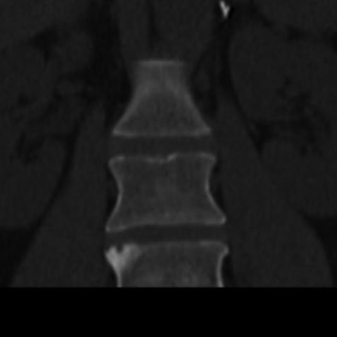
The main findings in the CT study were sclerosis, irregularities, and hyperostosis of the manubrium and medial ends of both clavicles (Fig. 1); asymmetric sacroiliitis (Fig. 2) and sclerosis and erosion of the superior-lateral corner of L4 vertebral body (Fig. 3).
Bone scintingraphy (Fig. 4) revealed intense radiotracer uptake in both clavicle medial ends and sternoclavicular joints. There was also radiotracer uptake in the sacroiliac joints (more pronounced on the right).
Discussion
SAPHO is an acronym that refers to an uncommon syndrome, composed of the combination of synovitis, acne, pustulosis, hyperostosis and osteitis. This entity was first described, less than 3 decades ago, by Chamot et al [1]. Due to its wide range of clinical presentations, it is considered, by some authors, a spectrum of diseases rather than a discrete syndrome [2].
It affects more often young adults, with a female preponderance.
The causes are unknown, although some authors support its integration in seronegative spondyloarthtropaties which is supported by the increasead prevalence of the HLA B27 allele, occasional presence of sacroiliitis, inflammatory bowel disease and psoriasis [3].
The most common dermatologic manifestations are palmoplantar pustulosis, severe acne, and psoriasis.
A fundamental component of SAPHO syndrome is an inflammatory osteitis, which corresponds, in the histopathologic study, to sterile neutrophilic pseudoabscesses [4]. The most frequent involved osteoarticular segment is the anterior thoracic wall, including sternoclavicular and sternocostal joints. Peripheral bones, spine and pelvic girdle can also be affected, with assimetric sacroiliitis being a common finding.
Characteristic radiographic findings include hyperostosis, which includes endosteal and periosteal proliferation and enthesopatic ossification. In association there are mixed areas of osteolysis. Adjacent joints show manifestations of arthritis, namely erosions and joint space narrowing. CT can depict these alterations in greater detail. MRI T2- weighted images with fat suppression or STIR can detect marrow oedema, which is useful to differentiate between chronic and acute lesions [3, 5]. Bone scintigraphy is a very sensitive modality, with radiotracer uptake present even before radiographic findings. A characteristic appearance is the “bull head” sign in the anterior chest wall, consisting of intense radiotracer uptake in sternoclavicular heads [6].
Radiologists must be aware of SAPHO spectrum disorders, given its characteristic clinical presentation and radiographic appearance and distribution, thus guiding appropriate therapeutic management.
Differential Diagnosis List
Final Diagnosis
SAPHO syndrome
Liscense
Figures
CT axial image

CT coronal image
Tc-99m methylene diphosphonate bone scintigraphy

CT axial image

Radiological Findings
Based on the provided CT and planar bone scintigraphy, the following key features can be observed:
1. Sclerotic changes and mild bone hyperplasia are visible in the pelvic region (including near the sacroiliac joints), with some areas showing local thickening or “hyperostosis,” suggesting a reactive bone process.
2. In the sternum, sternoclavicular joints, and certain vertebral bodies, there are imaging findings indicative of proliferative sclerosis or mixed bone lesions. CT may reveal localized bone thickening and possibly disorganized trabecular structure.
3. Bone scintigraphy shows high tracer uptake in the manubrium and sternoclavicular region, presenting a “bull’s head sign.” Additionally, increased uptake is noted in the sacroiliac joints of the pelvis, indicating active bone metabolism or osteitis-like changes.
Potential Diagnoses
- SAPHO Syndrome (Synovitis, Acne, Pustulosis, Hyperostosis, Osteitis): The patient presents with pustular skin lesions and polyarticular pain, along with radiological findings of osteoproliferative changes and inflammation in the sternoclavicular and sacroiliac joints. These findings align with SAPHO syndrome or its related spectrum.
- Axial Spondyloarthritis or Other Seronegative Spondyloarthropathies: Some patients may exhibit sacroiliitis and inflammatory changes in the spine, often accompanied by joint pain, but typically lack significant pustular skin involvement or marked sclerotic changes.
- Infectious Osteomyelitis: Although sclerosis or abnormal uptake can suggest infection, laboratory tests (e.g., WBC count, CRP) do not indicate a clear bacterial or fungal infection, and histology confirms the absence of cultivable pathogens.
Final Diagnosis
Considering the patient’s age, sex, cutaneous manifestations (pustular lesions), joint symptoms (polyarticular pain), and imaging findings (inflammatory and sclerotic changes in the sternoclavicular and sacroiliac joints, plus the “bull’s head sign” on bone scintigraphy), the most likely diagnosis is: SAPHO Syndrome.
If any doubt remains, further serological and imaging follow-up can be performed, and a bone biopsy may be considered to exclude rare conditions or confirm the absence of infectious inflammation.
Treatment and Rehabilitation Plan
1. Pharmacological Treatment:
· Use Non-Steroidal Anti-Inflammatory Drugs (NSAIDs) first to alleviate pain and joint swelling.
· For moderate to severe inflammation, consider short-term corticosteroids or immunomodulators (e.g., methotrexate, biologics) as assessed by a rheumatology specialist.
· For severe pustular lesions, add dermatological treatments (e.g., topical creams or oral retinoids), in collaboration with a dermatology specialist.
2. Physical Therapy and Rehabilitation:
· During the acute phase, avoid triggering or high-load exercises, focusing on pain and inflammation control, possibly with local heat application or physical modalities.
· In the chronic or remission phase, gradually initiate exercise programs targeting joint function and muscle endurance. Physical therapists or sports medicine specialists may guide the process, with goals including:
• Maintaining joint range of motion: gentle flexibility and ROM exercises;
• Strengthening the core and surrounding muscles: start with low-intensity resistance (elastic bands, light dumbbells), then progressively increase resistance and load;
• Improving cardiorespiratory endurance: engage in moderate-intensity aerobic activities such as brisk walking, cycling, or swimming, avoiding prolonged high-impact exercises to prevent excessive joint stress.
3. Applying the FITT-VP Principle:
· Frequency: Exercise 3-4 times per week, adjusting based on pain and fatigue.
· Intensity: Start low (e.g., 50%-60% of maximum heart rate) and gradually increase with symptom improvement and enhanced fitness.
· Time: 20-30 minutes per session, which can be extended to 45 minutes or more as tolerated.
· Type: Prefer low-impact aerobic exercises like cycling, swimming, or using an elliptical machine, combined with moderate-strength resistance training to maintain or enhance muscle strength.
· Progression: Regularly assess disease activity and joint function. If stable without new or worsening pain or swelling, gradually increase exercise duration or load.
· Volume/Plan: An initial goal may include at least 90-120 minutes weekly of cumulative aerobic exercise, 2-3 strength training sessions, and appropriate range-of-motion exercises.
4. Safety Considerations:
· Monitor symptom changes: If joint swelling or pain worsens significantly during exercise, stop and consult a specialist.
· Individual Assessment: Patients with osteoporosis or other comorbidities should undergo bone density and cardiopulmonary evaluations prior to starting a structured exercise program, ensuring safety and appropriateness.
Disclaimer: This report is intended solely for reference in medical analysis and does not replace in-person consultation or the guidance of a qualified physician. For specific diagnosis and treatment, please consult specialists in rheumatology, dermatology, and rehabilitation as needed.
Human Doctor Final Diagnosis
SAPHO syndrome