Medial clavicle fracture associated with arterial bleeding in the pectoralis major muscle


Clinical History
An 83-year-old man who takes Rivaroxaban presented at the emergency department after a bicycle accident. There was a hematoma around the medial clavicle and antalgic dysfunction of the left shoulder. Rapid expansion of the hematoma prompted an urgent Computed Tomography Angiography (CTA) of the thorax.
Imaging Findings
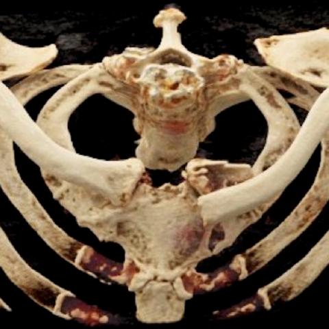
Conventional radiography showed a communitive, extra-articular fracture at the sternal end of the left clavicle. Non-contrast CT revealed an associated hematoma in the pectoralis major muscle and sternocleidomastoid muscle adjacent to the fracture site (Figures 1a, 1b). Subsequent CTA demonstrated a small focus of iodine contrast extravasation in the pectoralis major muscle (Figures 2a, 2b), in keeping with an arterial bleeding of one of the pectoral branches of the thoracoacromial artery. Significant volume increase of the hematoma was also causing progressive compression of the jugular vein.
Discussion
The majority of chest traumas are blunt injuries, which are related to chest wall injuries (e.g. fracture, hematoma) and pulmonary injury (e.g. pneumothorax, lung contusion), increasing patient morbidity and mortality [1]. The absence of bony thoracic injuries does not exclude other serious chest injuries such as a thoracic wall haemorrhage [2].
The clavicle connects the upper extremity to the trunk and protects the adjacent axillary and subclavicular neurovascular structures and lung apices. Vascular injuries are mostly seen with penetrating traumas, but rarely also occur in blunt traumas.
Clavicle fractures can be classified by the Allman classification. Fractures of the medial third are least frequent (2-6%), but are associated with chest trauma in up to 49% of cases and have the highest risk of associated neurovascular injuries. They are mostly seen in high-impact trauma [3,4]. The frequency of neurovascular injuries resulting from clavicle fractures is unknown, but the review of Mouzopoulos et al. discovered that 50% of subclavian artery injuries are found when the proximal clavicula is dislocated superiorly by traction of the sternocleidomastoid [5].
The typical presentation of a thoracic wall haemorrhage is a rapid-expanding mass as shown in multiple cases by Florescu et al. (2022) [6], typically within minutes to hours; however, delayed bleeding can occur after more than 24 hours [2,7]. In our patient, it was essential to be alert for this complication: arterial bleeding under anticoagulation has a high mortality rate. Thus, urgent imaging is needed. Chest CT angiography is the imaging tool of choice for stable patients, whereas catheter angiography is mandatory in unstable patients [6]. On CT, the location of the active bleeding point is typically seen as a small focus of iodine contrast extravasation in the arterial phase with dissemination in a delayed phase (e.g. after 65 seconds).
Thoracic wall arterial bleeding can either be treated by open exploration or by endovascular embolization [8]. Endovascular embolization has been proven successful in numerous cases for treatment of active bleeding [8–10]. Our patient was also successfully treated by selective embolization of a pectoral branch of the thoracoacromial artery.
In conclusion, rapid-progressive swelling of the thoracic wall should prompt additional imaging, even in absence of thoracic fractures. CTA is preferred imaging modality in stable patients. Medial clavicular fractures are associated with high-impact trauma and concomitant injuries. To our best knowledge, no previous case depicting bleeding of a thoracoacromial vessel following blunt trauma has been published.
Written informed patient consent for publication has been obtained.
Differential Diagnosis List
Final Diagnosis
Medial clavicular fracture complicated by arterial bleeding in the pectoralis major muscle
Liscense
This work is licensed under a Creative Commons Attribution-NonCommercial-ShareAlike 4.0 International License.
Figures


I. Imaging Findings
1. The local structure of the left medial segment of the clavicle (near the sternal end) appears irregular, suggesting a possible fracture or fracture line;
2. Noticeable soft tissue swelling in the adjacent area with increased density, indicating localized hematoma or blood infiltration signs;
3. Possible contrast extravasation in the arterial phase, suggesting active bleeding;
4. No obvious fracture signs detected in the rib sequence or the rest of the thoracic cage;
5. No apparent pneumothorax or large-scale parenchymal damage in the lung fields, and no evident mediastinal widening; however, carefully rule out potential hematoma involving mediastinal structures;
6. Other observed structures (such as the scapula, thoracic vertebrae, etc.) appear without significant morphological or structural abnormalities.
II. Potential Diagnoses
- Medial Clavicle Fracture with Arterial Injury
Imaging suggests a possible fracture at the medial end of the clavicle with significant soft tissue swelling and signs of active bleeding. The patient’s use of anticoagulant (Rivaroxaban) increases the risk of hemorrhage. Therefore, suspicion is high for a clavicle fracture with tearing or injury to nearby arterial branches (e.g., branches of the thoracoacromial artery) leading to bleeding. - Chest Wall Hematoma (Arterial Bleeding)
Rapidly progressing chest wall swelling could originate from a chest wall artery, including intercostal arteries or branches of the thoracoacromial artery. Arterial-phase contrast extravasation indicates bleeding from an arterial branch. Even without a clear fracture, this should be considered; however, a clavicle fracture or instability substantially raises the likelihood of vascular damage. - Other Vascular Lesions at the Cervicothoracic Junction (e.g., Pseudoaneurysm)
If the fracture segment or traumatic stimulus leads to arterial wall injury, a pseudoaneurysm or localized bulge could form. However, acute bleeding symptoms are more consistent with a sudden arterial disruption rather than a chronic pseudoaneurysm.
III. Final Diagnosis
Based on the patient’s history of trauma (bicycle accident), anticoagulant use, the suspected medial clavicle fracture on imaging, arterial-phase contrast extravasation in the soft tissues, and a rapidly progressing local hematoma, the findings most likely indicate:
"Left Medial Clavicle Fracture with Associated Injury and Bleeding of the Thoracoacromial Artery (or its branches)"
This corresponds with documented cases of high-energy trauma and medial clavicle fractures accompanied by vascular injury. Further assessment of the bleeding source or the extent of vascular damage may require interventional angiography or surgical exploration as indicated by clinical need.
IV. Treatment Plan and Rehabilitation Program
1. Treatment Strategy
- Emergency Hemostasis and Endovascular Treatment
– For patients with active arterial bleeding, urgent angiography and selective arterial embolization are recommended. If bleeding is significant or hematoma expansion is rapid and difficult to control, surgical exploration for hemostasis may be considered;
– Ongoing or adjusted anticoagulant therapy must be evaluated by specialists, balancing the patient’s underlying conditions with the current risk of bleeding. - Fracture Management
– If the clavicle fracture is significantly displaced or associated with vascular injury, an orthopedic consultation is warranted; open reduction and internal fixation (ORIF) may be required;
– If there is no severe displacement, conservative management (e.g., sling or figure-of-eight bandage) can be used with close monitoring of the wound, hematoma, and neurovascular status. - Supportive Treatment
– Pain management: Use analgesics and physical therapy measures to alleviate pain;
– Infection prevention: Prevent secondary infection of the hematoma;
– Cardiopulmonary function assessment: Especially in older patients, provide proper supportive therapy if needed.
2. Rehabilitation and Exercise Prescription
Rehabilitation training should account for the advanced age of the patient, chest trauma, and possible osteoporosis, progressing gradually to avoid excessive traction.
- Early Stage (within 1–2 weeks post-surgery or acute phase)
– Main goals: Alleviate pain, prevent joint stiffness;
– Mode of exercise: Passive or assisted active movements of the shoulder and elbow joints, avoiding large ranges of motion;
– Frequency: 2–3 times a day, 5–10 minutes each time, adjusted according to pain tolerance. - Mid Stage (2–6 weeks)
– Main goals: Promote fracture healing, maintain muscle strength, and gradually restore range of motion;
– Mode of exercise: Within the limits of pain control, progressively increase shoulder mobility exercises and muscle strengthening (e.g., light resistance with elastic bands);
– Frequency: 3–4 times a week, 15–20 minutes each session, appropriately increasing resistance and range of motion. - Late Stage (after 6 weeks)
– Main goals: Strengthen the shoulder girdle and chest muscles, improve upper limb function;
– Mode of exercise: Gradually add moderate-intensity upper limb resistance exercises, functional training simulating daily activities;
– Frequency: 3–5 times a week, 20–30 minutes each session, progressing under the guidance of a physician or therapist according to fracture healing status.
Throughout the rehabilitation process, monitor for pain, swelling, and neurovascular symptoms. If any abnormality occurs (e.g., hematoma expansion, severe pain), seek immediate medical advice or contact the attending physician.
Disclaimer:
This report is a reference analysis based on current imaging and clinical information and does not substitute for in-person consultation or a certified medical professional’s diagnosis and treatment advice. If you have any questions or if your condition changes, please seek medical attention promptly.
Human Doctor Final Diagnosis
Medial clavicular fracture complicated by arterial bleeding in the pectoralis major muscle