Van Buchem disease


Clinical History
A 46-year-old female patient with cervical pain, cephalea, hypoacusis and decrease in visual acuity.
Imaging Findings
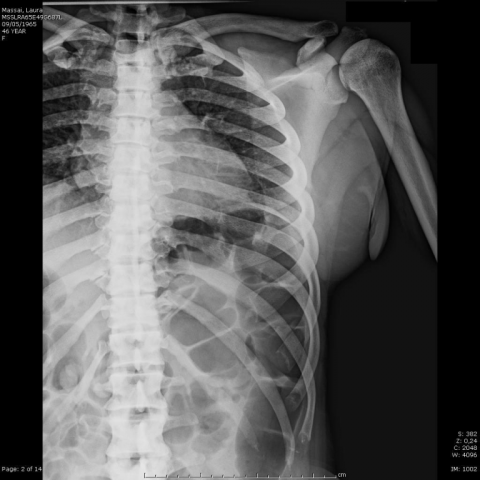
A 46-year-old female patient came to our attention with cervical pain, hypoacusis, cephalea and decrease in visual acuity. Cervical spine RX was performed, showing gross thickening of the calvarium, particularly in the frontal and basi-occipital regions, bone overgrowth of jaw, hyperostosis of cervical vertebrae (Fig. 1). Skeleton RX demonstrates significant hyperostosis of clavicle and ribs (Fig. 3), cortical thickening of long bones diaphyses with reduction of medullary canal, especially in femur and tibia (Fig. 4). Also the pelvis appears thickened. Bodies of thoracic and lumbar vertebrae are less involved than spinous processes (Fig. 2).
CT confirms frontal and occipital thickening (>3 cm), petrous bone hyperostosis and reduction of internal and external auditory canal (Fig. 5).
Laboratory shows increase in alkaline phosphatase and bone densitometry (BMD) values are significantly elevated (2.050 g/cm³), T-score 11, 8. Laboratory, clinical and radiological features are strongly suggestive of Van Buchem disease.
Discussion
Van Buchem disease (VBD) is a rare autosomal recessive bone pathology, classified as familiar generalised cortical hyperostosis (FGCH) and characterised by cortical endosteal bone tissue overgrowth.
Two forms of FGCH are known.
Type 1 or Van Buchem disease, with autosomal recessive transmission. Bone abnormalities begin in the first decade and are progressive, alkaline phosphatase (ALP) levels are elevated.
Type 2 or Worth disease, with autosomal dominant transmission. In this pathology the bone overgrowth stops in second decade with normal ALP value [6].
Pathogenesis is related to increase in osteoblasts activity. VBD is determined by deletion in non-coding sequence of SOST gene. The SOST gene, localised in chromosome 17q12-q21, encode sclerostin, which inhibit bone growth. In VBD this protein is absent or significantly low [3-4; 7-8].
Many bones are affected by Hyperostosis: jaws and skull in first, ribs, clavicle and long bones diaphysis. Bodies of vertebras are normal, posterior arches are involved instead. Anomalies are symmetric, progressive determining skeleton overweight up to three times.
Diagnosis is based on familiar anamnesis and radiological features; laboratory reveals ALP high values. Histology shows normal bone structure.
RX shows thickening of the calvarium, particularly in the frontal and basi-occipital regions. Skull base and petrous bones are also involved, sometimes paranasal sinus obliteration is observed. Jaw is a typical localisation, with increase of sagittal and transverse diameters, condyles thickening and open gonial angle [1-2]. Long bones show cortical hyperostosis of the dyaphyses and medullary canal reduction. Hand and feet bones are thickened also at epiphyses. Ribs and clavicle presents significant hyperostosis, cervical column results more involved than dorsal and lumbar column. Pelvis is less hyperostotic than other regions [9-10].
CT confirms RX evidences and is useful in the study of petrous bone, cranial and peripheral nerves foramina.
MR is helpful in evaluation of pheriperal nerves pathology.
Neurological issues are frequent in VBD as nerve encroachment, paralysis of peripheal nerves (V, VII, VIII, X), hypoacusis and hearing loss by sclerosis of ossicular chain and pyramidal region of petrous bone, optic atrophy by stricture of optic foramen. Intracranial hypertension symptoms, cognitive and motor deficit are less common. No case of bone fractures are reported [1-2; 9-10].
Differential diagnosis is between Albers-Schönberg disease, in which skull and jaw are not typically involved, vertebrae are hyperostotic, incidence of fractures is elevated [2], and Engelmann-Camurati disease, characterised by skull, long bones and pelvis cortical thickening (jaw is not affected), pain at extremities, muscular fatigue and typical waddling gait [5].
Differential Diagnosis List
Final Diagnosis
Van Buchem disease
Liscense
Figures
RX skull


RX column and pelvis


RX ribs and clavicles

RX long bones


CT images


Imaging Findings
In this case, the patient (female, 46 years old) primarily underwent X-ray and partial CT examinations of the skull, vertebrae, pelvis, and long bones of the extremities. A comprehensive review suggests the following features:
- Skull and Skull Base: Noticeable thickening of the skull, especially in the frontal region and the basi-occipital area; the calvarium appears widened, and both petrous bones are involved. Some patients may exhibit sclerosis of the ossicles and narrowing of the external/internal auditory canals.
- Mandible: Thickening of the mandibular body, angle, and condyle, indicating cortical hyperostosis in the jaw area, potentially altering the occlusal relationship.
- Spine: Marked thickening of the posterior elements in the cervical and thoracic vertebrae, with the vertebral body shape largely normal; however, the lamina at the back is significantly thickened, which may lead to relative narrowing of the spinal canal.
- Thorax and Ribs: The ribs show varying degrees of cortical thickening, and the thoracic skeletal structure overall has a tendency toward thickening.
- Pelvis and Long Bones: The pelvis shows comparatively mild thickening, but cortical thickening is still visible in the ilium and ischium; the long bones of the extremities (femur, tibia, humerus, etc.) also reveal noticeable cortical thickening with narrowed medullary cavities.
Laboratory tests typically show elevated serum alkaline phosphatase (ALP), though this report is based solely on imaging findings and references to previous literature. The patient’s symptoms include neck pain, headaches, hearing loss, and reduced vision, all potentially related to nerve impingement caused by narrowed cranial foramina.
Possible Diagnoses
Based on the patient’s clinical presentations (hearing impairment, vision loss, neck pain, headaches) and imaging findings (widespread cortical thickening, prominent involvement of the skull and mandible), the following differential diagnoses are considered:
- Van Buchem Disease (Familial Exuberant Osteosclerosis, Type 1):
- A rare autosomal recessive disorder characterized by significant cortical bone thickening, commonly seen in the jaw, skull base, and diaphyses of long bones.
- Typical findings include progressive skull thickening, elevated serum alkaline phosphatase, and symptoms related to nerve compression (e.g., hearing and vision impairment).
- Albers-Schönberg Disease (Osteopetrosis or Marble Bone Disease):
- Bone sclerosis typically involves the vertebrae, pelvis, and long bones, with a propensity for fractures; the skull and mandible are not commonly thickened.
- Can be inherited in either an autosomal dominant or recessive pattern and is often accompanied by limited bone marrow space.
- Engelmann-Camurati Disease (Progressive Diaphyseal Dysplasia):
- Primarily involves cortical thickening of the long bones and skull, though jaw involvement is less characteristic; patients often present with limb pain, muscle weakness, and a waddling gait.
Final Diagnosis
Taking into account the patient’s age, symptoms (neck pain, headaches, hearing and vision impairment), imaging findings (notably significant cortical thickening in the mandible and skull base, thickening of the thoracic posterior elements), hereditary factors, and frequently elevated serum alkaline phosphatase in such conditions, the most likely diagnosis is Van Buchem Disease (Type 1 Familial Exuberant Osteosclerosis).
For further confirmation, one may consider:
- Genetic testing (related to mutations or deletions in the SOST gene).
- Blood tests (including ALP, calcium and phosphate metabolism, and other bone biomarkers).
- MRI of suspected nerve compression sites to assess the extent of neural involvement.
Treatment Plan and Rehabilitation
1. Treatment Strategies
- Conservative Management: There is currently no specific medication to reverse bone thickening, and treatment mainly involves symptomatic relief such as analgesics for neck pain and headaches, or neurotrophic agents to protect compressed nerves.
- Surgical Intervention: Decompression surgery may be considered if neural compression becomes severe (e.g., causing irreversible damage to the optic or auditory nerves). Orthognathic surgery may be evaluated if mandibular overgrowth significantly affects occlusion.
- Assistive Devices: Patients with hearing impairment can be fitted with hearing aids, while those with vision impairment should receive regular ophthalmological follow-up or consider optical correction.
2. Rehabilitation/Exercise Prescription and Recommendations
Given the nature of Van Buchem Disease with increased bone density, the risk of fracture is not typically elevated as in other bone diseases. However, thickened bones can cause nerve compression and limit physical movement. Therefore, the following points should be noted in rehabilitation:
- Frequency: Recommend 3-5 sessions of rehabilitation or moderate exercise per week, spread over different days, avoiding continuous high-intensity workouts.
- Intensity: Focus on low to moderate intensity, avoiding high-impact exercises that place excessive stress on the cervical spine and vertebrae. Low-impact aerobic activities (such as stationary cycling or elliptical machines) or pool therapy are good options.
- Time: Each session should last about 30-45 minutes, including 5-10 minutes for warm-up and 5 minutes for cool-down.
- Type: Prefer exercises that place minimal strain on joints, such as swimming, stationary biking, Tai Chi, or gentle stretching routines. Avoid positions that could exacerbate cervical or cranial pressure.
- Progression: Gradually increase from low to moderate intensity, while monitoring neck pain and neurological symptoms. If headaches worsen or if hearing or vision further declines, discontinue and seek medical evaluation promptly.
Given that the patient already experiences headaches, hearing loss, and reduced vision, any exercise should be performed under professional guidance, with caution regarding excessive movement of the head and neck to minimize the risk of nerve injury. If the patient’s cardiopulmonary function is adequate, a long-term, comprehensive plan can be developed under the guidance of both physicians and rehabilitation specialists, incorporating muscle strength, flexibility, and balance training.
Disclaimer
This report provides a reference analysis based on available patient information and imaging data and does not replace an in-person consultation or professional medical advice. Specific treatment and rehabilitation plans should be formulated and adjusted in real-time by specialized physicians and rehabilitation therapists according to the patient’s actual condition.
Human Doctor Final Diagnosis
Van Buchem disease