Monostotic fibrous dysplasia of the mandible


Clinical History
An 11-year-old healthy girl presented with some discomfort over the right mandible. There was no history of trauma, constitutional symptoms or recent surgery. Clinical examination and blood tests were unremarkable. Plain radiograph was requested by the maxillofacial surgeon as he was concerned regarding a bone tumour.
Imaging Findings
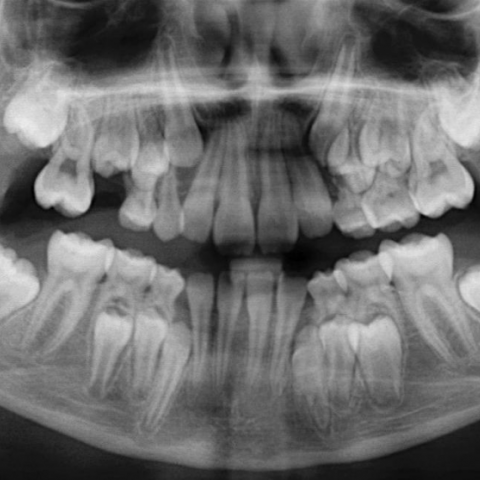
Plain radiograph (Fig. 1) showed subtle loss of cortico-medullary differentiation and increased lucency affecting the right ramus of the mandible. Subsequent CT (Fig. 2) showed ground-glass change and expansion of the right ramus of the mandible extending to the mandibular condyle. No aggressive periosteal reaction was seen and appearances were in keeping with a benign bone lesion. Follow-up CT (Fig. 3) was performed in 12 months time on the request of surgeons which showed similar ground-glass appearance and no associated soft tissue mass. The patient remained symptoms free during this period, therefore biopsy was not considered.
Discussion
Fibrous dysplasia (FD) is a benign fibro-osseous developmental anomaly of the mesenchymal precursor of bone, predominantly seen in 1st and 2nd decades of life with 75% of patients present before the age of 30 [1, 2] with about equal incidence in males and females [3]. FD becomes dormant by the third decade. Skull base and face is involved in 25% of cases, most commonly affecting maxilla and mandible, followed by frontal bone.
Histopathologically, FD is composed of fibrous tissue containing bone trabeculae [3].
Mandibular FD may be asymptomatic, or patients present with non-specific symptoms including pain, swelling, tenderness, and stress or overt fracture [1, 2].
FD on plain radiograph has a ground-glass or smoky matrix with altered and remodelled normal bone architecture. The lesions are mostly medulla based, may be eccentric within the affected bone, but not cortical. Ultimately, the affected bone undergoes an expansile remodelling secondary to the enlarging mass of fibro-osseous tissue. On CT, lesions typically have ground-glass appearance with sclerotic well-defined borders and no aggressive periosteal reaction. MRI is rather nonspecific and lesions appear homogeneous/ mildly heterogeneous marrow lesions, mainly hyperintense to fat on T2W, and hypointense to muscle on T1W images.
FD may undergo malignant transformation in 0.5% cases, most commonly osteosarcoma, followed by fibrosarcoma and chondrosarcoma [4].
Differentials include ossifying fibroma (OF), chronic recurrent multifocal osteomyelitis (CRMO) and Paget's disease. OF typically has thick, bony rim and lower density centre and occurs mostly in the third and fourth decades of life. OF tends to occur more commonly in the anterior mandible and is smaller in size, whereas fibrous dysplasia is more common in the posterior mandible and the lesions tend to be larger. CRMO manifest as chronic mandibular pain and/or swelling. Initial stages demonstrate a lytic process with associated variable amounts of sclerosis and associated soft-tissue inflammation, without evidence of discrete abscess formation. Mandibular disease has been shown to be associated with lesions in other skull or facial bones such as the maxilla and zygoma [5].
Treatment of FD involves bony recontouring for cosmesis and function however, the lesions can show surprising growth potential during their active growth phase if treated surgically [6]. Considering this fact and self-limiting nature of the lesion, the patient was treated conservatively.
Radiological features of FD of the mandible have been described. Familiarity with these features would help to reassure patients and alleviate their anxiety.
Differential Diagnosis List
Final Diagnosis
Fibrous dysplasia of the mandible
Liscense
Figures
Mandible plain X-ray
CT of the mandible


Coronal and Axial CT of the mandible


Medical Imaging Analysis Report
I. Imaging Findings
1. From the panoramic X-ray, the right mandible shows a slight expansion in morphology, with a “ground-glass” appearance of the bone. No obvious cortical discontinuity is observed.
2. Axial and coronal CT scans indicate heterogeneous bone density in the affected region of the mandible. “Smoky” or “ground-glass” density changes are visible, primarily within the medullary portion. The boundaries are relatively clear, and no significant erosion or destructive features are noted.
3. No evident soft tissue mass or extra-osseous extension is observed, nor any signs of root resorption or widened periodontal spaces.
4. The surrounding soft tissue structures appear essentially normal, with no notable pathological lymph node enlargement.
II. Potential Diagnoses
- Fibrous Dysplasia (FD)
- Commonly occurs in adolescents and young adults, often between the ages of 10 and 20, which matches this patient’s age.
- Radiographically, it typically shows a “ground-glass” or “smoky” appearance, with lesions primarily in the medullary region.
- The lesion can grow expansively, but there is usually no significant cortical destruction.
- Ossifying Fibroma (OF)
- More frequently seen in the third or fourth decades of life. Once mature, a well-defined thick sclerotic rim is typically visible.
- OF may share some radiological similarities with fibrous dysplasia, but OF often presents with a more localized sclerotic boundary and is commonly found in the anterior jaw.
- Chronic Recurrent Multifocal Osteomyelitis (CRMO)
- May present with chronic pain or swelling of the mandible. Imaging can show lytic lesions with irregular sclerosis.
- Often accompanied by changes in other craniofacial bones, with or without soft tissue inflammatory signs.
- Paget’s Disease
- Most commonly seen in middle-aged or elderly individuals. The affected bones may exhibit enlargement, deformity, and alternating areas of sclerosis and lysis.
- This patient is relatively young and lacks typical radiographic changes; hence, Paget’s disease is less likely.
III. Final Diagnosis
Given that the patient is an 11-year-old female with minimal clinical symptoms, normal routine blood tests, characteristic “ground-glass” changes on imaging, and expansile bony proliferation, the most likely diagnosis is Fibrous Dysplasia (FD).
This condition generally becomes more stable after adolescence. In the absence of significant functional impairment or cosmetic concerns, regular follow-up may be sufficient. If further clarification is required, a biopsy or consultation with a pathology department can be considered.
IV. Treatment Plan and Rehabilitation
1. Treatment Strategy:
(1) Conservative Observation: Since fibrous dysplasia often stabilizes after adolescence or early adulthood, and because this patient’s clinical symptoms are mild, regular follow-up—including imaging and clinical assessments—can be considered.
(2) Surgical Treatment: If severe occlusal dysfunction, significant pain, or cosmetic concerns arise, curettage or remodeling surgery of the affected bone may be considered. Surgical intervention is generally recommended once skeletal growth is more stable, reducing the chance of additional procedures caused by ongoing lesion growth.
(3) Pain Management: For persistent pain, use of analgesics or localized treatment measures under specialist guidance may be considered.
2. Rehabilitation and Exercise Prescription:
(1) General Principles: This condition usually has limited impact on daily activities; however, attention should be paid to skeletal integrity, particularly in the mandible, to avoid intense impacts or activities that could cause facial injury.
(2) FITT-VP Principle:
• Frequency: 3-5 sessions of moderate exercise per week. Choose low-impact aerobic activities (e.g., walking, swimming, cycling) based on the patient’s age and physical condition.
• Intensity: Mild to moderate, avoiding overly strenuous workouts. Adjust according to heart rate or perceived exertion, keeping within tolerable limits of daily activity.
• Time: Around 20-30 minutes per session, with duration modified according to the patient’s energy levels and growth status.
• Type: Emphasize safe, low-impact exercises. Activities involving potential head or facial trauma should be avoided or approached with caution. If playing sports, appropriate facial protection is advised.
• Progression: Increase exercise time and intensity gradually if there are no adverse symptoms, avoiding abrupt changes.
• Volume & Progression: Taking into account the patient’s daily routine and interests, increase total exercise volume or duration by about 10-15% per week.
(3) Special Precautions:
• Protect the mandible if its structure is weakened, minimizing risk of injury.
• If increased pain, marked swelling, or functional difficulties occur, reduce activity and seek medical reassessment.
Disclaimer:
This report is based on the currently available information and is provided for reference purposes only. It cannot replace an in-person consultation or the diagnosis and treatment advice of a qualified medical professional. The detailed therapeutic plan should be determined by combining clinical manifestations, individual differences, and professional medical assessment.
Human Doctor Final Diagnosis
Fibrous dysplasia of the mandible