Thrombosed persistent median artery - A rare cause of carpal tunnel syndrome


Clinical History
A 36-year-old male software professional presented with complaints of pain in the left wrist and lateral half of the hand for 20 days. The patient suggests worsening of pain during activity. No history of trauma was elicited. He was referred for ultrasound of the left wrist to assess the median nerve.
Imaging Findings
On ultrasound the median nerve appears unremarkable. Axial images of distal forearm depict a hypoechoic rounded structure adjacent to the median nerve. On sagittal sections it corresponds to a tubular hypoechoic structure extending from the forearm into the carpal tunnel. This characteristic course coupled with arterial flow pattern on Doppler imaging clinches the diagnosis of a persistent median artery (PMA).Within the carpal tunnel this artery appears distended with echogenic contents and shows no colour flow confirming thrombosis.
Contrast enhanced MR angiography depicts an additional artery in the forearm between the radial and ulnar arteries originating from the anterior interosseous artery and extending into the carpal tunnel confirming a PMA. Absence of normal flow void and hyperintense signal within the artery on T1W/PD fat saturated images and rim enhancement with a central filling defect on contrast enhanced images confirm thrombosis of the PMA within the carpal tunnel. The thrombosed PMA is seen indenting the median nerve.
Discussion
Carpal tunnel syndrome (CTS) is the most common entrapment neuropathy of the upper extremity and occurs due to compression of the median nerve within the carpal tunnel. Various idiopathic, traumatic, inflammatory, infective, endocrine and congenital conditions as well as mass lesions (eg, ganglion cyst, neurogenic tumours) can cause CTS [1].
The median artery, a branch of the axial artery is the dominant blood supply to the developing hand in early fetal life and normally involutes before birth.
A persistent median artery (PMA) is one of the rare congenital conditions implicated as a cause of CTS [2]. The approximate incidence of PMA as a cause of CTS is 6% [3]. A persistent median artery (PMA) is commonly a branch of one of the forearm arteries and courses through the carpal tunnel, usually adjacent to the median nerve [4]. Thrombosis of the PMA within the carpal tunnel probably occurs due to repeated stress and vibration [5]. A thrombosed PMA likely causes CTS due to mass effect on the median nerve and possible irritation of the nerve by inflammatory changes surrounding the area of thrombosis. The cause of thrombosis in our patient, a software professional, was probably prolonged dorsiflexion at the wrist during work.
Ultrasound and MRI of the wrist are commonly requested in suspected cases of CTS. Apart from assessing the median nerve, a diligent search for abnormal structures occupying the carpal tunnel should be made in all cases. It is imperative to trace any abnormal structure within the carpal tunnel proximally, to assess possible extension into the forearm. A tubular structure in the forearm extending into the carpal tunnel separate from the median nerve is suggestive of a PMA with colour Doppler and contrast enhanced MR angiography aiding in confirmation [6]. A bifid median nerve (two nerve bundles within a common perineurium) in the carpal tunnel is a common association with a PMA and can be easily be easily detected on ultrasound to avoid inadvertent injury during surgery [7].
Treatment of a thrombosed PMA involves medical therapy to aid thrombolysis with surgery reserved for failed cases. Release of the transverse carpal ligament and resection of the artery are possible surgical options [2, 8]. Before resection it is essential to make sure that the PMA does not substantially contribute to hand circulation. Contrast enhanced MR angiography offers a relatively non-invasive means of visualising the extent of involvement of the artery in forming the palmar arch.
Differential Diagnosis List
Final Diagnosis
Thrombosed persistent median artery causing carpal tunnel syndrome.
Liscense
Figures
Colour Doppler and Spectral Doppler


T1Weighted MRI


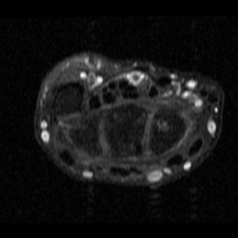
Proton Density Weighted fat saturated MRI


Post contrast T1 Weighted fat saturated MRI

Reformatted coronal MIP image of contrast enhanced MR angiography

High frequency ultrasound of wrist (10 to 16 MHz)


I. Imaging Findings
1. Ultrasound Examination: On the palmar side of the left wrist, adjacent to the median nerve, a tubular structure is visible, showing anechoic lumen with local blood flow signals. A low-echo area suggestive of thrombus formation is noted, indicating possible lumen narrowing with heterogeneous echoes. The median nerve from the proximal transverse carpal ligament to the carpal tunnel segment appears thickened with increased cross-sectional area, consistent with common findings of Carpal Tunnel Syndrome (CTS).
2. MRI Examination: Axial images of the wrist reveal an enlarged median nerve within the carpal tunnel, with T2WI showing high signal intensity changes. A tubular vascular structure is seen adjacent to the median nerve, correlating with ultrasound findings. Contrast-enhanced images demonstrate this tubular structure is connected to the forearm arterial system, suggesting it could be a Persistent Median Artery (PMA). Some sequences show low-signal filling suggestive of thrombus formation.
II. Potential Diagnoses
- 1. Carpal Tunnel Syndrome (CTS)
This arises due to compression of the median nerve in the carpal tunnel, causing pain, numbness, and sensory disturbances. Common causes include bony hypertrophy, tendon sheath thickening, trauma, or other space-occupying lesions. In this case, the patient’s job involves prolonged wrist extension or flexion (computer software work), which may trigger or worsen compression of the median nerve within the carpal tunnel. - 2. Carpal Tunnel Occupying Lesion Caused by Persistent Median Artery
A Persistent Median Artery results from the embryonic median artery failing to regress. In most cases, it does not cause clinical symptoms. However, if a thrombus forms or if the artery is abnormally thickened within the carpal tunnel, it can compress the median nerve, leading to CTS symptoms. The imaging findings here indicate a tubular artery structure with signs of thrombosis, suggesting a mass effect or inflammatory irritation. - 3. Other Lesions in the Carpal Tunnel
Although the main presentation in this case is consistent with CTS caused by a PMA, other rarer conditions such as local tumors, tendon or tendon sheath inflammation, and nerve sheath tumors should also be considered for differential diagnosis.
III. Final Diagnosis
Considering the patient’s occupational factors (sedentary work, prolonged fixed wrist posture), clinical symptoms (left wrist pain, exacerbation with activity, sensory disturbances in the median nerve distribution), and imaging findings (thickened median nerve within the carpal tunnel, adjacent persistent median artery with thrombosis), the most likely final diagnosis is “Carpal Tunnel Syndrome (CTS) caused by a thrombosed Persistent Median Artery.”
IV. Treatment Plan and Rehabilitation Program
Based on the above diagnosis, the following treatment and rehabilitation recommendations are provided:
-
Conservative Treatment and Medication
• Non-steroidal anti-inflammatory drugs (NSAIDs) can be used to reduce local pain and inflammation.
• Wearing a wrist splint (brace) to minimize pressure in the carpal tunnel, especially at night or during long periods of desk work.
• If thrombosis is confirmed, consider local or systemic anticoagulation/thrombolytic therapy under specialist guidance, to observe recanalization of the artery and symptom relief. -
Surgical Treatment
• If conservative treatment fails or symptoms are severe and significantly affect daily life, consider carpal tunnel release surgery to relieve pressure on the median nerve.
• For a Persistent Median Artery with a significant thrombus and if it is confirmed not to be a major contributor to hand blood supply, consultation with both vascular surgery and hand surgery specialists may lead to arterial resection or vascular intervention. -
Rehabilitation and Exercise Prescription
Following the FITT-VP principle (Frequency, Intensity, Time, Type, Volume, Progression), the recommendations are as follows:
• Frequency: 3–5 times per week.
• Intensity: Keep exercises at a light to moderate level; stop immediately if any significant discomfort appears and adjust intensity accordingly.
• Time: 20–30 minutes per session, possibly divided into multiple shorter sessions.
• Type: Focus on gentle flexion and extension exercises of the wrist, isometric strengthening of the forearm muscles, and low-impact aerobic exercises like walking or using an elliptical machine to improve overall circulation.
• Progression: As symptoms improve, gradually add wrist stabilization exercises, such as squeezing a grip ball or using small dumbbells or resistance bands, but proceed slowly and carefully.
• Individualization: If the patient has comorbidities (hypertension, diabetes, etc.), exercise intensity and methods should be adjusted under professional medical or rehabilitation guidance.
During the rehabilitation period, it is crucial to avoid maintaining the wrist in a prolonged hyperextended or flexed position. Take regular breaks for wrist rest and gentle stretching to reduce continuous compression risk.
Disclaimer: This report is based on the provided medical history and imaging data for reference only and does not replace an in-person consultation or a professional doctor's diagnosis and treatment advice. If you have any questions or changes in your condition, please consult a specialist and undergo further examinations in a timely manner.
Human Doctor Final Diagnosis
Thrombosed persistent median artery causing carpal tunnel syndrome.