Parsonage Turner syndrome as a cause of fatty atrophy of multiple shoulder muscles


Clinical History
A 45-year-old female patient presented with pain, numbness and weakness involving the right shoulder and arm for the past 6 months. The left upper and bilateral lower limbs were normal. No history of trauma or antecedent history of infection was elicited.
Imaging Findings
The supraspinatus, infraspinatus, subscapularis, teres minor and deltoid muscles show reduced muscle bulk and abnormal T1W hyperintense signal suggestive of fatty replacement. The tendons of these muscles retain their normal hypointense signal. The pectoralis muscles and the muscles of the upper arm show normal muscle bulk and signal. No definite rotator cuff tendon tears nor features of impingement were seen. No abnormality was noted in the suprascapular notch, spinoglenoid notch and the quadrilateral space (not shown) where possible compression of the suprascapular nerve and axillary nerve may occur. Imaging of the cervical spine revealed no significant spinal cord compression or neural foraminal narrowing.
Discussion
Parsonage Turner syndrome (PTS) is characterised by severe pain and weakness involving at least one muscle in the shoulder. This condition has also been otherwise described including ‘acute brachial neuritis’ and neuralgic amyotrophy [1, 2]. It often mimics other common conditions like cervical spondylosis, rotator cuff pathology and shoulder impingement, which may result in delayed diagnosis and inappropriate treatment [1]. Infectious and immunologic processes including prior viral infections and vaccination are thought to play a role in the pathogenesis of brachial neuritis [1, 2].
The common clinical features in PTS include severe sudden-onset pain and weakness. Sensory symptoms like hypaesthesia and paraesthesia and rarely autonomic symptoms like skin changes, nail changes and increased sweating in affected areas have also been described [3]. The disorder is usually self-limiting with most patients showing varying degrees of improvement months to years after onset [1].
MRI is the ideal imaging modality for visualising the muscle changes secondary to denervation caused by brachial neuritis. Undesrstanding of the innervation of the various muscles around the shoulder is essential in assessing patterns of involvement on MRI. The supraspinatus and infraspinatus muscles are supplied by the suprascapular nerve, the subscapularis muscle by the subscapular nerve and the deltoid and teres minor muscles by the axillary nerve. The commonest nerve involved is the suprascapular nerve with the supraspinatus and infraspinatus muscles being the most commonly-affected muscles [1, 2]. The signal changes occurring within muscles on MRI vary based on the phase of the disease [2]. A diffuse increase in T2W signal within muscles is seen in the acute phase of denervation. In the subacute and chronic phases changes of atrophy and fatty infiltration occur, visualised as increased signal on T1W images.
Our case showed reduced muscle bulk and significant fatty replacement of the supraspinatus, infraspinatus, subscapularis, teres minor and deltoid muscles corresponding to involvement of the suprascapular, subscapular and axillary nerves. Involvement of these three nerves was described in one case in the study by Gaskin and Helms [1]. Our case showed no abnormality in the region of the suprascapular/ spinoglenoid notch or quadrilateral space (not shown) where possible compression of the suprascapular and axillary nerve may occur. The cervical spine showed no significant abnormality. No history of chronic disuse of right shoulder prior to onset of symptoms was elicited. The chronic nature of the patient’s symptoms coupled with the characteristic imaging findings favoured the diagnosis of chronic phase of PTS.
Differential Diagnosis List
Final Diagnosis
Chronic phase of Parsonage Turner syndrome
Liscense
Figures
Axial T1W MRI Right shoulder


Coronal T2W MRI Right shoulder

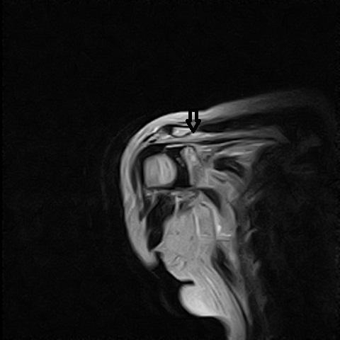

Sagittal T2W MRI Cervical spine

Medical Imaging Analysis Report
I. Imaging Findings
The patient is a 45-year-old female complaining of right shoulder and upper arm pain, numbness, and weakness for 6 months. Imaging (shoulder MRI) primarily shows:
- Significant atrophy and fatty infiltration of the supraspinatus, infraspinatus, subscapularis, teres minor, and deltoid muscles, appearing as hyperintense signal changes on T1-weighted images.
- The distribution of lesions corresponds to the muscle groups around the shoulder joint, with innervation by the suprascapular nerve, subscapular nerve, and axillary nerve.
- No apparent compressive lesions in the suprascapular notch, spinoglenoid notch, or quadrilateral space; cervical MRI shows no obvious nerve root or spinal canal stenosis or notable compression.
- No definitive signs of chronic disuse changes (e.g., localized inactivity in a certain region), suggesting an imaging presentation more consistent with a neurogenic etiology.
II. Potential Diagnoses
Based on the sudden onset of severe shoulder pain followed by muscle weakness and atrophy, together with the imaging findings, possible diagnoses or differential diagnoses include:
- Parsonage-Turner Syndrome (acute brachial neuritis)
Features: acute or subacute onset, severe shoulder pain, followed by weakness and atrophy of the involved muscle groups. Imaging can show neurogenic changes in the shoulder region. This fits the case scenario. - Rotator Cuff Tear or Subacromial Impingement Syndrome
Features: often related to trauma, overuse, or degenerative changes, primarily affecting the rotator cuff tendons. However, MRI typically shows tendon tears or rupture signs, which does not completely align with the extensive involvement of multiple muscle groups in this case. - Cervical Radiculopathy
Features: cervical disc herniation or spinal canal stenosis causing nerve root compression can lead to weakness, pain, or sensory disturbances at the corresponding segment, but cervical MRI in this case does not demonstrate clear evidence of nerve root compression. - Other Peripheral Nerve Lesions
Such as local suprascapular or axillary nerve compression. However, there is no sign of local anatomical narrowing or mass effect in this case.
III. Most Likely Final Diagnosis
Based on the patient’s symptoms (acute onset of pain followed by muscle weakness, atrophy, and sensory disturbances), imaging characteristics (neurogenic atrophy of multiple shoulder muscles, fatty infiltration, no evident local compression or cervical radiculopathy), and history (around six months of persistent course, not due to trauma or chronic disuse), the most likely diagnosis is:
Chronic phase Parsonage-Turner Syndrome (late stage of acute brachial neuritis).
IV. Treatment Plan and Rehabilitation
After confirming the diagnosis, the following treatment and rehabilitation plan may be considered:
- Conservative Treatment
- During the acute and painful phases, nonsteroidal anti-inflammatory drugs (NSAIDs) or local analgesics can be used to alleviate pain.
- In cases of severe pain, short-term corticosteroid therapy under specialist guidance, combined with neurotrophic agents, may be considered.
- Physical Therapy and Rehabilitation Exercises
- Initial Rehabilitation Phase (Pain Control Stage)
Local heat application and gentle passive range-of-motion exercises are recommended, avoiding excessive loading. Gentle activities can be performed 2–3 times per day, 5–10 minutes each session. - Gradual Progression Phase (Functional Recovery Stage)
After pain subsides, gradually introduce active shoulder movement with elastic band or mild resistance exercises. According to tolerance, train 3–5 times per week, 20–30 minutes each session, focusing on strengthening shoulder abduction, internal rotation, and external rotation. - Later Strengthening Phase (Muscle Strength Enhancement)
As muscle strength recovers, resistance or load can be gradually increased, along with exercises to improve shoulder stability, such as dynamic shoulder exercises in a supported position. Perform 3–5 times per week, at least 30 minutes each time, emphasizing joint symmetry and posture correction.
- Initial Rehabilitation Phase (Pain Control Stage)
- Surgical or Interventional Treatment
- Most acute brachial neuritis cases improve with conservative treatment and rehabilitation, and generally do not require surgery.
- If severe nerve entrapment or other structural abnormalities are suspected, further imaging evaluation or surgical exploration may be warranted.
- Other Considerations
- Because this condition may involve sensory deficits in the innervated regions, caution should be taken to avoid strain or excessive joint movements during exercises and to maintain shoulder stability.
- In patients with other comorbidities (e.g., osteoporosis, compromised cardiopulmonary function), the intensity and frequency of exercises should be reduced accordingly and closely monitored.
Disclaimer: This report is for medical analysis reference only and does not replace in-person clinical consultation or professional medical advice. Specific treatment plans should be based on the patient’s actual situation and be determined by qualified clinical physicians.
Human Doctor Final Diagnosis
Chronic phase of Parsonage Turner syndrome