Dilatation of basivertebral veins mimicking osteoblastic lesions on contrast enhanced CT.


Clinical History
A 56-year-old man with a history of left pneumonectomy for suspicion of a tumour of the left upper and lower pulmonary lobe, with post-operative histology disproving neoplasia and being positive for fibrotic tissue with chronic inflammation signs, presented with thoracic back pain without neurological deficit. PSA level was normal.
Imaging Findings
A contrast enhanced thoraco-abdominal CT was performed demonstrating left brachiocephalic vein stenosis due to left mediastinal deviation post left pneumonectomy with collateral vessels of the left chest wall. In addition, it showed sclerotic lesions with symmetric localisation at the posterior part of the vertebral bodies T2 to T7, corresponding to basivertebral vein distribution.
On MRI the sclerotic lesions at the posterior part of the vertebral bodies T2 to T7 described at the CT were not found, especially on T1 weighted sequences that are very sensitive to sclerotic lesions.
Furthermore, a contrast-enhanced cervical CT (covering the upper thoracic spine) with early acquisition time effectuated just a few days later showed no evidence of sclerotic vertebral lesions at the same locations.
Discussion
Pneumonectomy is the treatment of choice for bronchogenic carcinoma and intractable end-stage lung diseases [1]. It involves reasonable anatomic changes and a number of potential complications concerning the respiratory system, the cardiovascular system, and the pleural space [2]. In the post pneumonectomy period, the mediastinum either remains stationary or gradually shifts toward the postpneumonectomy space as a result of hyperextension of the remaining lung [1]. After left pneumonectomy, the heart rotates counterclockwise into the vacant left pleural space [3].
In the above case, left brachiocephalic vein stenosis occurred in the post-operative period due to the left mediastinal deviation post pneumonectomy. As a result a collateral venous circulation via the vertebral pathway and left chest wall veins was developed. In brachiocephalic vein stenosis there are the following options for the development of a collateral circulation: 1) Collateral flow through deep and superficial veins of the back, chest, and neck into the contralateral jugular, subclavian, and brachiocephalic veins or 2) Collateral flow through superficial chest wall veins such as the internal mammary and intercostal veins into the azygos (if right-sided occlusion) or hemiazygos (if left-sided occlusion) or into inferior epigastric veins [4].
In this case, there is development of left chest wall collaterals with high venous pressure leading to opacification of the dilated basivertebral veins. As a result there is an increase in vertebral density at the posterior middle part of T2 to T7 vertebrae that was mistaken for osteoblastic lesions at the contrast enhanced CT [5]. However the symmetry and location of the sclerotic lesions should raise suspicion of basivertebral vein opacification. MRI excluded the diagnosis of pathologic vertebral lesions. A contrast-enhanced cervical CT (including the upper dorsal spine), performed just a few days after the initial thoraco-abdominal contrast enhanced CT, showed no evidence of osteoblastic vertebral lesions because of the absence of contrast in the basivertebral veins, due to earlier acquisition time post contrast injection.
Our case illustrates that contrast opacification of dilated basivertebral veins can lead to false diagnosis of bone metastases. To our knowledge there has been only one case previously reported in the literature with focal vertebral marrow enhancement due to basivertebral dilatation after superior vena cava obstruction [5].
Differential Diagnosis List
Final Diagnosis
Dilatation of basivertebral veins due to brachiocephalic vein stenosis postpneumonectomy.
Liscense
Figures
Contrast enhanced CT


MRI


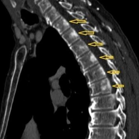
Sagittal contrast enhanced CT

Axial contrast enhanced CT image with MIP reconstruction

Imaging Findings
On enhanced chest CT scans, the patient shows:
- Significant postoperative changes in the left thoracic cage; the left lung has been removed and there is a noticeable cavity in the left thoracic cavity.
- Mediastinal shift to the left, with evidence of local stenosis in the left brachiocephalic vein (left innominate vein).
- Between the T2 and T7 vertebral bodies in the central posterior region, contrast agent enhancement is visible, resembling proliferative changes of vertebral trabeculae. This can be easily confused with sclerotic changes of bone metastases, but the distribution is symmetrical and follows the trajectory of the vertebral basal veins.
- MRI shows normal signal in the corresponding vertebrae with no signs of space-occupying lesions or bone destruction, ruling out pathological bone changes.
- A repeat contrast-enhanced (neck and chest) CT in an earlier phase showed no enhancement in this part of the vertebral body, indicating that the previously observed enhancement was actually contrast within the vessels.
- Compensatory dilatation of the veins in the left thoracic wall suggests the possibility of collateral circulation formation.
Potential Diagnoses
Based on the imaging findings and the patient's history, possible diagnoses include:
- Bone metastases: Since the patient underwent lung surgery, and there appears to be suspicious sclerotic lesions in the vertebrae on imaging, bone metastases must be considered. However, the patient’s PSA level is normal, MRI has ruled out bone destruction and a solid tumor lesion, and the lesions are symmetric and vascular in appearance, which is atypical for metastatic disease.
- Pseud–sclerotic changes due to dilatation of the vertebral basal veins: Stenosis of the left brachiocephalic vein impairs venous return, leading to collateral circulation and dilatation of the vertebral basal veins. On CT, the retained contrast in these veins may appear as “sclerotic” lesions, but there is no actual bone destruction or neoplastic process.
- Inflammatory vertebral lesions (e.g., vertebral hemangioma or other vascular proliferations): While these can present as local density or signal changes, they are often focal or patchy, with distinct MRI features. Further examinations do not support such a diagnosis in this case.
Final Diagnosis
Taking into account the patient’s left lung pneumonectomy, mediastinal shift, stenosis of the left brachiocephalic vein, and the emergence of significant collateral venous circulation, the conclusion is:
Most likely diagnosis: “Pseud–sclerotic changes arising from dilatation of the vertebral basal veins caused by stenosis of the left brachiocephalic vein,” rather than bone metastases.
MRI clearly excludes true bone lesions, and the variable contrast enhancement of the basal veins in different CT phases further supports this conclusion.
Treatment Plan and Rehabilitation
First, a comprehensive evaluation of the left brachiocephalic (innominate) vein stenosis and potential vascular flow obstruction is necessary. Consultation with vascular surgeons or cardiothoracic surgeons may be required to consider the following options:
- Conservative management: If the patient’s symptoms are mild and there are no signs of severe venous obstruction, clinical observation and regular follow-up imaging are recommended. Venotonics or medications that improve venous return and protect the vascular endothelium can be considered.
- Vascular intervention or surgical treatment: For significant venous return impairment or severe symptoms, options such as stent placement or surgical correction may be considered to relieve the stenosis.
- Symptomatic treatment: In cases of local pain or discomfort, appropriate analgesics may be used. If there is associated back muscle tension or postural issues, physiotherapy can be added for relief.
An individualized plan should be established for rehabilitation and functional training:
- FITT-VP principle (Frequency, Intensity, Time, Type, Volume, Progression):
- Frequency: Exercise 3–5 times per week, starting with fewer sessions for 1–2 weeks to monitor tolerance, then potentially increasing to 5 sessions per week.
- Intensity: Engage in low-to-moderate intensity aerobic activities (e.g., walking on flat ground, slow cycling), gradually increasing intensity as cardiopulmonary fitness improves.
- Time: Begin with sessions of 15–20 minutes and increase by 5 minutes weekly as tolerated, aiming for 30–45 minutes over time.
- Type: Emphasize full-body aerobic exercise, supplementing with simple core strengthening and shoulder/back stretching exercises. Avoid heavy lifting or high-impact activities.
- Volume and Progression: Adjust according to the patient’s physical capacity, pain level, and venous return. If significant relief of back pain and good cardiopulmonary endurance are observed, consider increasing walking speed or adding low-load resistance training.
- For patients with reduced lung function or lower cardiopulmonary capacity, integrate specialized breathing exercises and endurance training, such as:
- Basic breathing exercises: Diaphragmatic breathing, pursed-lip breathing, etc., to improve lung capacity and breathing efficiency.
- Light aerobic exercise combined with breathing training: For instance, deep breathing during walking or cycling to enhance oxygen exchange.
- Monitor closely for symptoms like chest pain, dyspnea, or marked engorgement of head and neck veins. Should these symptoms worsen significantly, discontinue activity and seek medical advice promptly.
In summary, this patient should follow a low-risk aerobic endurance program combined with local muscle-strengthening exercises, progressing gradually. Long-term monitoring of symptoms and imaging findings is essential.
Disclaimer: This report is for reference only and does not replace in-person consultation or professional medical guidance. If you experience any discomfort or a worsening of symptoms, please seek medical attention or consult a qualified healthcare professional promptly.
Human Doctor Final Diagnosis
Dilatation of basivertebral veins due to brachiocephalic vein stenosis postpneumonectomy.