Intraosseous lipoma of the talus


Clinical History
In January 2013, this patient presented with pain and swelling in the left ankle after a twisting injury six days ago. Her bloods were normal.
Imaging Findings
Figure 1. AP radio-graph shows a well-corticated 14mm round-to-ovoid lucent lesion within the head/neck of the talus. No fracture is demonstrated. No cortical breach or periosteal reaction is demonstrated.
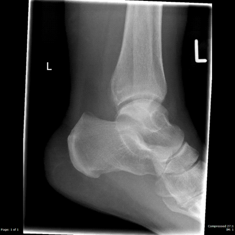
Figure 2. Lateral radio-graph shows a well-corticated 14mm round-to-ovoid lucent lesion within the head/neck of the talus. The lesion does not reach the articular surface of the talus, excluding an OCD. No fracture is demonstrated. No cortical breach or periosteal reaction is demonstrated.
Figure 3. MRI TI Sag sequences shows a well defined ovoid lesion within the talus. The lesion shows a high signal on T1 sequences.
Figure 4. MRI PD FS sequence shows signal loss within the lesion, indicating that the lesion is of fat density, hence narrowing the differential.
Discussion
Differential Diagnosis:
Based on Radiographs: (FOG-MACHINES)...there is a wide differential for solitary bone lesions.
Fibrous Dysplasia
Osteoblastoma
Giant Cell Tumour
Metastasis / Myeloma
Aneurysmal Bone Cyst
Chondroblastoma / Chondromyxoid Fibroma
Hyperparathyroidism (brown tumours) / Haemangioma
Infection
Non-ossifying Fibroma
Eosinophilic Granuloma / Enchondroma
Solitary Bone Cyst
Given the appearance and location of the lesion, more realistic possibilities include:
Simple Bone Cyst
Fibrous Dysplasia
Enchondroma
Chondromyxoid Fibroma
Based on MRI scans:
Lipoma
Liposarcoma.
This lesion is an intra-osseous lipoma. These are rare, benign tumours with an incidence of <0.1% per year. They are usually asymptomatic and diagnosed incidentally. Most patients are middle aged with a male predominance. The calcaneum is the most commonly affected site in the foot. Cases within the talus are rare.
Intraosseous lipomas have 3 stages: (1) Purely radio-lucent with cortical expansion. (2) Similar radio-graphic appearance but contains localised calcification. (3) Lesion shows ossification around the calcified fat of the outer rim.
Teaching points:
There is a wide differential for solitary bone lesions, however some of them may have distinguishing features e.g. an associated clinical history or associated clinical finding which helps to narrow the diagnosis, such as a raised white cell count and pyrexia for infection. Some of the lesions are also more common at certain sites e.g. a chondroblastoma more commonly occurs in the epiphysis. Others have distinctive features e.g. fibrous dysplasia has a ground glass appearance. Certain tumours are also more typical in certain age groups e.g. a chondroblastoma is more common in the 1st-2nd decade. Intra-osseous lipomas can also mimic an OCD when occurring close/adjacent to the articular surface of the talus.
Further investigation with MRI is sometimes necessary to narrow the differential diagnosis. The loss of signal on the STIR sequence indicated that the lesion contained fat. The most common fat based lesion occurring in the calcaneum is a lipoma. These are rare within the talus but cases have been reported. (Please see references). The lack of aggressive features on both radiographs and MRI makes this lesion less likely to represent a liposarcoma, however malignant transformation of a lipoma to a liposarcoma has been reported and repeat imaging is advised if the patient develops new clinical symptoms.
Differential Diagnosis List
Final Diagnosis
Intraosseous lipoma of the talus
Liscense
Figures
Plain radiography

Plain radiography
MR

MR

1. Imaging Findings
Based on the provided X-ray and MRI of the left foot and ankle joint, there is a well-defined lucent lesion within the talus. The local cortical bone shows slight expansion but no significant destruction or obvious soft tissue swelling. The lesion appears more radiolucent on the X-ray with some demarcation from the surrounding bone. On MRI sequences (especially STIR), the lesion primarily exhibits fat signal characteristics, which is attenuated on fat suppression or STIR sequences, suggesting possible fatty components. No obvious bone marrow edema or significant soft tissue abnormalities are observed around the lesion; likewise, there is no clear cartilage defect or subchondral bone plate disruption at the adjacent articular surface, and no obvious joint effusion is noted. Overall, the imaging findings appear benign.
2. Potential Diagnoses
Considering the patient’s age, clinical presentation, and imaging features, the differential diagnoses include:
- Simple Bone Cyst: Common in adolescents, typically presents as a radiolucent bone lesion. However, this case involves a middle-aged patient, and the MRI shows significant fatty components, which does not fully match a simple bone cyst.
- Fibrous Dysplasia: Often exhibits a “ground-glass” appearance and usually lacks prominent fat signals, which does not fit this case.
- Cartilaginous Lesions (e.g., Enchondroma, Chondromyxoid Fibroma): On X-ray, these may show calcifications or cartilage matrix signals. However, the predominantly fatty signal on MRI in this case is not typical for cartilaginous lesions.
- Intraosseous Lipoma: A radiolucent area with an internal fat signal is commonly found in the calcaneus but can rarely appear in the talus. The MRI findings here indicate a fatty component, consistent with this diagnosis.
- Liposarcoma: A highly malignant tumor that can involve bone destruction or soft tissue invasion. MRI may show heterogeneous fat signals with possible enhancement. However, there are no apparent aggressive features in this case, suggesting a benign lesion.
3. Final Diagnosis
Given the patient’s demographic (middle-aged female), imaging studies (especially MRI indicating a fat-dominant lesion without obvious malignant features), and clinical symptoms (pain in the dorsal foot and ankle region post-sprain), the most likely diagnosis is Intraosseous Lipoma. While malignant transformation rates are extremely low, the rare possibility of liposarcoma should be kept in mind. If symptoms worsen or follow-up imaging shows significant progression, further evaluation or biopsy may be warranted.
4. Treatment Plan and Rehabilitation Program
Under the current clinical and imaging context, most intraosseous lipomas can be managed conservatively unless there is persistent pain, a risk of pathological fracture, or suspicious malignant features. Main strategies include:
- Conservative Observation: Regular follow-up and imaging (X-ray or MRI) to monitor for lesion progression or new symptoms.
- Surgical Intervention: In cases of recurrent pain or suspected malignant change, consider curettage of the lesion with bone grafting or internal fixation to stabilize the joint structure.
Rehabilitation and Exercise Prescription Recommendations:
According to the FITT-VP principle (Frequency, Intensity, Time, Type, Progression), the specific recommendations are:
- Early Recovery: While pain and swelling persist, use low or non-weight-bearing exercises, such as seated or supine ankle dorsiflexion and plantarflexion. Perform 2–3 times per day at low to moderate intensity, avoiding significant pain.
- Intermediate Training: Gradually increase standing and walking time, introducing mild ankle weight-bearing exercises such as partial squats or resistance band exercises for the ankle. Keep the intensity moderate, 3–5 sessions per week, 20–30 minutes each.
- Advanced Progression: As alignment stabilizes and symptoms resolve, incorporate workouts that enhance flexibility and strength, such as light jogging or single-leg balance exercises. Carefully control intensity and watch for ankle discomfort, increasing training volume and difficulty gradually.
- Safety Precautions: If the patient is at risk for osteoporosis or other factors, reduce the loading and exercise intensity accordingly. If severe pain or swelling arises during recovery, stop activities and seek medical attention promptly.
In summary, maintaining regular follow-ups, moderate rehabilitation exercises, and monitoring lesion changes is the main management approach. If there is rapid lesion enlargement, structural instability, or significant pain, surgical intervention or further imaging and pathological evaluation should be considered.
Disclaimer: This report is based on the provided medical history and imaging data for reference only and does not replace an in-person consultation or specialist opinion. Should there be further questions or changes in symptoms, please seek timely medical advice or specialist consultation.
Human Doctor Final Diagnosis
Intraosseous lipoma of the talus