Chronic ALPSA with recurrent shoulder instability


Clinical History
Recurrent right shoulder instability in a 20-year-old man with a history of previous shoulder dislocation.
Imaging Findings
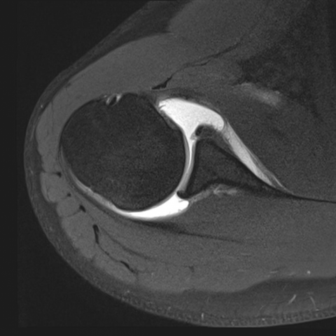
The anterior glenoid rim is bare, with the detached and medially displaced anterior labrum seen as a low-signal mass attached to the scapular neck by the periosteal sleeve. The labrum also rotates inferiorly, which makes it appear large on axial images and inferior to the scapular neck on the coronal images. A small contrast-filled crease is visible on the axial images where the lateral margin of the detached synovium-covered labrum meets the glenoid rim, which can be identified on arthroscopy.
Note that the anterior band of the inferior glenohumeral ligament attaches onto the ALPSA lesion (Figure 1), in part accounting for the instability. A subtle Hill-Sachs lesion is also observed at the posterior superior humeral head (Figure 1).
Findings are consistent with chronic anterior labrum periosteal sleeve avulsion (ALSPA).
Discussion
Anterior labrum periosteal sleeve avulsion (ALPSA) lesion is common in chronic instability of the shoulder, especially in those with repeated dislocations. It is considered a variant of anteroinferior labral tear and consists of a tear of the anteroinferior labrum with associated capsuloperiosteal stripping [1]. It can originate from a Perthes lesion, when continued traction transmitted along the anterior band of the inferior glenohumeral ligament strips the periosteum which remains attached to the labrum medially along the glenoid neck. The anteroinferior labrum retracts further medially with each episode of dislocation or subluxation and becomes adherent to the glenoid neck through scarred tissue [2].
ALPSA is one of the manifestations of chronic glenohumeral instability which affects the inferior labral-ligametous complex, the primary passive stabilizer of the joint. Affected individuals may develop shoulder pain and recurrent dislocations following initial traumatic dislocation. MRI allows lesion characterisation for the purpose of treatment planning, with the primary goal of stabilisation.
In chronic ALPSA lesions, the torn and medially displaced anteroinferior labrum is seen along the glenoid neck as a low signal mass, representing the scarred down labrum held by the periosteal sleeve. It is often rotated inferiorly and appears larger on axial images, with abnormal thickening and flattening resulting from healing with synovial fibrous tissue between the labrum and glenoid margin. A small cleft or separation can be observed between the glenoid margin and the labrum. The anteriorinferior glenoid rim is bare or deficient [1-2].
Differential Diagnosis List
Final Diagnosis
Chronic anterior labrum periosteal sleeve avulsion (ALPSA)
Liscense
Figures
T1W FS MR arthrogram axial image
T2W FS MR arthrogram coronal oblique image

Sagittal T1W FS MR arthrogram image

Imaging Findings
1. MRI shows a significant tear in the anteroinferior region of the right shoulder labrum, with visible medial retraction of the labrum to the neck of the glenoid.
2. The torn anteroinferior labrum is “pulled” and adheres to the glenoid neck by the preserved periosteal sleeve, seen as low-signal soft tissue at the corresponding site, suggesting scar formation.
3. The labrum appears abnormally thickened, with a small gap-like signal (fissure) between the labrum and the glenoid rim; the anteroinferior glenoid rim appears relatively flattened or slightly deficient.
4. Considering the patient’s history (repeated shoulder subluxation or dislocation) and imaging findings, these are consistent with ALPSA (Anterior Labral Periosteal Sleeve Avulsion) changes in the context of chronic shoulder instability.
Potential Diagnoses
- ALPSA (Anterior Labral Periosteal Sleeve Avulsion) Lesion
Cause: Related to repeated dislocations, involving a tear of the anteroinferior labrum along with stripping of the periosteal sleeve and scar fixation; the chronic shoulder instability and noted medial displacement of the anteroinferior labrum on imaging match the key points of this diagnosis. - Bankart Lesion
Cause: Also occurs at the anteroinferior labrum and is a common lesion in traumatic shoulder instability; however, a standard Bankart tear typically does not exhibit significant medial displacement, often presenting as a simpler tear. - Perthes Lesion
Cause: Similar to Bankart, but the periosteum and anteroinferior labrum attachment remain intact, with the labrum still connected to the periosteum; if the avulsion progresses or is repeatedly pulled, it can evolve into an ALPSA lesion.
Final Diagnosis
Considering the patient’s age (20 years), multiple shoulder dislocations, and MRI findings of an anteroinferior labrum that is displaced medially and attached to the periosteal sleeve, the most likely diagnosis is: An anterior labral periosteal sleeve avulsion (ALPSA) lesion.
Treatment Plan and Rehabilitation
1. Surgical Treatment: For patients with multiple dislocations, higher degrees of instability, or significantly limited range of motion, arthroscopic debridement and labral repair (e.g., using suture anchors) can effectively restore joint stability and reduce the risk of re-dislocation.
2. Conservative Treatment: If dislocations are less severe and relatively infrequent, conservative management may be chosen, including appropriate immobilization (e.g., shoulder brace) and rehabilitation exercises, avoiding large-amplitude abduction and external rotation movements.
3. Rehabilitation Principles (FITT-VP): Gradual functional recovery training is required after either surgical or conservative treatment. It is generally divided into the following phases:
- Frequency: Start at 2–3 times per week in the early stage, and gradually increase to 3–5 times per week based on recovery.
- Intensity: Begin with low resistance and short duration exercises, avoiding excessive force and high-stress joint positions, and gradually increase abduction and external rotation range of motion.
- Time: About 20–30 minutes per session, adjusted according to the patient’s tolerance.
- Type: Includes passive range of motion exercises, strengthening of muscles around the shoulder (such as the rotator cuff), and gradually transitions to active resistance training.
- Volume & Progression: As pain subsides and range of motion improves, increase load, difficulty, and duration under the guidance of specialists or physical therapists, ensuring proper technique and safety.
4. Exercise Prescription Examples:
- Early Stage: Passive external and internal rotation, and abduction exercises with the elbow flexed, twice daily, 10–15 repetitions each time, with smooth, controlled movements.
- Mid Stage: Add resistance band exercises for external and internal rotation, 2–3 times per week, 2–3 sets per session, 10–15 repetitions per set.
- Late Stage: Incorporate functional exercises such as front and lateral raises with dumbbells, strengthening the rotator cuff and deltoid muscles.
- Focus on core strength and scapular stability exercises (e.g., push-ups, supine press, and scapular retraction) to ensure overall shoulder stability.
Safety Reminder: If significant pain, joint clicking, or discomfort occurs during training, stop immediately and seek medical evaluation to prevent secondary injuries or recurrent dislocation.
Disclaimer: This report is for reference only and cannot replace an in-person consultation or professional medical advice. Patients should develop final diagnosis and rehabilitation plans based on their individual conditions and the guidance of a specialist.
Human Doctor Final Diagnosis
Chronic anterior labrum periosteal sleeve avulsion (ALPSA)