Maffucci syndrome


Clinical History
A 44-year-old woman with a known skeletal disorder since her childhood presented with chest and shoulder pain.
Physical examination revealed hand deformities with several bluish nodules of hard consistency, multiple swellings scattered in several ribs and difficulty in mobilizing the shoulders.
Imaging Findings
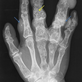
Plain radiographs revealed features compatible with S. Maffucci in the hands (Fig. 1), ribs, scapula and humerus (Fig. 2).
Enchondromas are radiolucent lesions with osseous expansile remodelling (yellow arrows) and vascular malformations in the adjacent soft tissues have phleboliths (blue arrows).
Discussion
Maffucci Syndrome is a rare mesodermal dysplasia, which is characterized by multiple enchondromas (Ollier disease) with co-existing vascular malformations [1].
It is a non-hereditary congenital disease and the diagnosis is made early in life. It has no gender predominance and affected individuals have normal stature and normal intelligence.
Enchondromas affect short tubular bones, occurring predominantly in the hands (87%) and less frequently in ribs (31%), humerus (42%) and scapula (25%) [2, 3].
It carries a significant risk of fracture and malignant transformation (23%) [1], mostly to chondrosarcoma, which is higher than in Ollier disease and significantly higher than in solitary endochroma of tubular bones. Other malignant tumours have been associated with this syndrome, such as glioma, ovarian and pancreatic cancer, and for that reason long-term follow-up evaluations are necessary.
The classification of vascular anomalies has been reviewed by the International Society for the Study of Vascular Anomalies (ISSVA) [4] in order to unify the nomenclature in this matter. According to this classification, there are two main types of vascular anomalies, vascular tumours and vascular malformations; the differentiation is based on clinical appearance, radiological and pathological features and biological behaviour. Vascular tumours, the most common being infantile haemangioma, represent a neoplastic growth of vascular endothelial cells, and can be benign, locally aggressive/borderline or malignant. In contrast, vascular malformations are considered to be vascular structural anomalies with no neoplastic proliferation of endothelial cells. The latter never disappears and tends to increase with age.
Vascular malformations are further subdivided in low-flow (capillary, venous or lymphatic), high-flow (arterial malformation, arteriovenous fistula or arteriovenous malformation) and combined, according to haemodynamic features and on predominant anomalous channels. This differentiation is quite important, since their morbidity and also their management are entirely different (arterial embolization for high-flow and sclerotherapy for low-flow malformations) [5].
Low-flow malformations are the most common type of vascular malformation and the best indicator is the presence of phleboliths, typically seen as calcified nodules, which is the case of Maffucci syndrome: a enchondromatosis associated with low-flow venous malformations[5].
In plain radiographs, enchondromas are characteristic and computed tomography and magnetic resonance imaging are helpful when malignant degeneration is suspected.
Vascular malformations are well diagnosed on the basis of clinical history and physical findings, but colour Doppler ultrasound and magnetic resonance imaging can be used to characterize these lesions.
No medical care is needed in asymptomatic patients and surgery is indicated for special cases [4].
Differential Diagnosis List
Final Diagnosis
Maffucci syndrome
Liscense
Figures
Hands

Chest

Medical Analysis Report
1. Radiological Findings
From the provided hand and chest X-ray images, the following findings can be observed:
• Multiple radiolucent bone lesions in the hands, primarily affecting the metacarpals and phalanges. Some lesions show mild expansile changes, and scattered small patchy or spot-like dense shadows are seen (possibly indicating calcification or phleboliths).
• Chest X-ray shows multiple expansile bony lesions involving both shoulder girdles (including the proximal humerus and scapula) and multiple ribs.
• Calcific foci within the soft tissues suggestive of venous malformations or so-called “phleboliths,” correlating with previously described “blue nodules.”
• Overall radiological features are consistent with cartilaginous-origin bone lesions coexisting with low-flow vascular malformations (venous malformations).
2. Possible Diagnoses
- Multiple Enchondromatosis (Ollier Disease): Characterized by multiple enchondromas. However, it typically lacks significant vascular malformations, so simple multiple enchondromas would be in doubt if vascular anomalies are present.
- Maffucci Syndrome: Based on multiple enchondromas (similar to Ollier disease), combined with low-flow venous malformations or cavernous hemangiomas. Subcutaneous or soft tissue “blue nodules” and phleboliths are commonly noted. The morphological presentation and clinical symptoms in this case are quite typical.
- Chondrosarcoma or Malignant Transformation: Multiple enchondromas carry a certain risk of malignant change. Significant bone destruction, a thick rim of calcification, or a soft tissue mass may indicate malignant transformation. Further CT, MRI, or biopsy can help confirm or rule it out.
Given the early onset, multiple cartilaginous lesions, and prominent low-flow vascular malformations, Maffucci Syndrome is most likely. If any lesion shows rapid enlargement or severe structural destruction, one must be cautious about possible malignant transformation (e.g., chondrosarcoma).
3. Final Diagnosis
Considering the radiological characteristics, the patient’s history of bone disease since childhood, and the clinical presentation, the most likely diagnosis is: Maffucci Syndrome.
If there is any concern about malignancy or suspicious changes (rapid growth, persistent and worsening pain, swelling in soft tissue, etc.), further MRI and pathological biopsy are recommended to rule out chondrosarcoma or other malignant conditions.
4. Treatment Plan and Rehabilitation
According to current literature and clinical practice, patients with Maffucci Syndrome who have no symptoms or only mild symptoms are usually managed with observation and regular follow-up. Surgical intervention is considered only when there is significant pain, functional impairment, or suspicion of malignancy. Specific recommendations are as follows:
- Conservative Management:
- Regular imaging follow-up (X-ray or MRI) to monitor lesion growth and check for signs of malignant transformation.
- If low-flow vascular malformations cause pain or cosmetic issues, consider sclerotherapy in vascular surgery or interventional radiology to reduce the size of the malformation.
- Surgical Treatment:
- For severe deformities causing functional impairment, pathologic fractures, or large enchondromas, procedures such as corrective surgery, curettage with bone grafting, and internal fixation may be required.
- If malignancy is suspected, a biopsy should be obtained promptly, and tumor resection surgery may be considered depending on the diagnosis.
Rehabilitation and Exercise Prescription Recommendations:
Since the patient’s limbs and thorax are affected, it is important to consider bone fragility and joint mobility during exercise. It is recommended to proceed under the guidance of a specialist in rehabilitation, gradually implementing:
- Type of Exercise: Low-impact activities (e.g., Tai Chi, yoga, light swimming, or walking in water) are recommended. Avoid high-intensity or collision-prone sports.
- Frequency: 3–5 times per week, adjusted according to joint flexibility and pain levels.
- Intensity: From low to moderate, avoiding severe pain or obvious discomfort. Use subjective exertion ratings (RPE) at a “somewhat hard” level.
- Time: 20–30 minutes each time, which can be divided into segments based on tolerance.
- Type (Mode of Exercise): Prefer activities with minimal joint loading that enhance blood circulation, such as active joint mobilization, stretching, and flexibility exercises to maintain and improve range of motion.
- Progression: After 2–4 weeks of consistent training, you may gradually increase the duration or frequency of exercise based on the patient’s tolerance.
- Precautions: If significant pain, swelling, or suspected fracture appears, stop the movement immediately and seek medical evaluation. Special caution is needed in areas with possible vascular malformations to avoid abrupt impacts.
Disclaimer:
This analysis report is for reference only and cannot replace an in-person consultation or the diagnosis and treatment advice of a professional physician. If you have any questions or changes in condition, please seek medical attention promptly and follow the guidance of a specialist doctor.
Human Doctor Final Diagnosis
Maffucci syndrome