Plexiform neurofibroma with limb hypertrophy


Clinical History
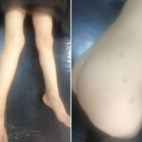
A 4-year-old boy presented for right lower limb mass with lower limbs discrepancy. The mass increased gradually in size causing intermittent throbbing pain. Physical examination showed a solid, mobile, mildly tender mass of the right calf. The boy and his brother have multiple pigmented macules (Fig. 1).
Imaging Findings
On X-ray the right leg is longer than the left one. Minimal bowing of the right tibial and fibular shafts was noted, as well as an increased soft tissue density. (Fig. 2)
MRI showed a soft tissue mass of the posterior compartment of the right leg. The mass was infiltrating, showing lobulated margins and extending from the distal thigh and popliteal fossa to the plantar aspect of the foot. It demonstrated an intermediate signal on T1-weighted images, a high signal on T2 and minimal enhancement after gadolinium administration. Axial T2-images showed multiple target appearances within the mass. (Fig. 3-4)
Discussion
Neurofibromatosis is an autosomal dominant multisystem genetic disorder, resulting from deletion/mutation of the NF1 gene responsible of production of a tumour suppressor protein: "neurofibromin". Lack of neurofibromin induces malformations of the skin, skeleton and nervous system. The prevalence of clinically diagnosed cases ranges from 1/2000 to 1/5000. [1]
The skeleton is commonly involved in NF1: spinal deformities, sphenoid and tibial bone dysplasia, excessive bone and soft-tissue growth. [2]
Dysplasia of long bones, most frequently the tibia, is seen in 7.1% of cases. [2]
In general it presents as tibial bowing with cortical thinning, increased risk of fracture and pseudarthrosis. In such cases limb discrepancy results from shortening of the affected limb, however, limb overgrowth may occur without dysplasia as in our case where the long limb demonstrates a soft tissue mass. It is postulated that the tumour-associated hyperaemia somehow induces limb overgrowth. [3]
The diagnosis of NF is clinical. [4] It is made in our case by the presence of three of seven clinical criteria (cafe-au-lait spots, tibial dysplasia and a first degree relative with cafe-au-lait spots); this clinical context and the above described MRI findings lead to the diagnosis of plexiform neurofibroma (PN).
PN is pathognomonic of NF1, found in 27% of cases. [5] It is an ill-defined infiltrating mass involving the nerve, muscles and adjacent fat, consequently often non totally resectable. [6] PN typically involves the trunk and extremities but also may affect the head, neck, urinary bladder or even the mesentery. [7]
On MRI it presents as an infiltrative mass of fascicular aspect, with hypo/iso T1 signal, iso/hyper T2 signal with subtle or no enhancement after gadolinium administration. A characteristic MR finding is the ”target sign” on axial T2 images: a low signal centre of collagen fibres and a high signal rim of myxomatous material. [8]
Besides the clinical picture, MRI is the diagnostic modality of PN. Angio-CT or MR also help assess the vascularization for any pre-surgical embolisation. [9]
Clinical context is the most important clue for diagnosis. When large, the neurofibroma causes hypertrophy of the affected member giving a presentation called “elephantiasis neuromatosa”. [1]
Differential diagnoses include proteus syndrome, a sporadic disease of tissue and bone with limb overgrowth, vascular malformations and muscular asymmetry [11, 12] and Kasabach-Merritt syndrome that is thrombocytopenia with large infiltrating soft tissue haemangiomas. [13] The first syndrome is unlikely since the patient's disease is familial, the second includes bright T2 significantly enhancing lesions unlike our case.
Differential Diagnosis List
Final Diagnosis
Plexiform neurofibroma
Liscense
Figures
Photograph of the trunk and lower limbs

X-ray of both legs

MRI of both legs

MRI of both legs

1. Imaging Findings
Based on the patient’s X-ray and MRI images, the following observations are noted:
- A soft tissue mass in the right lower leg with relatively unclear boundaries, showing an infiltrative growth pattern, causing certain bony changes. The X-ray indicates that the right tibia is slightly longer than the contralateral side and mildly curved. This correlates with the clinically observed mild overgrowth of the affected limb.
- The MRI demonstrates that on T1-weighted images, the mass appears as low or iso-intense signal, while on T2-weighted images, it shows high or mixed signal intensity. A local “target sign” (relatively low signal in the center and high signal on the periphery) suggests the presence of collagen fibers and myxoid matrix.
- The lesion extends primarily around the nerve bundles, also involving surrounding soft tissue with irregular margins, consistent with the typical imaging features of a plexiform neurofibroma.
2. Potential Diagnoses
Based on the above imaging findings and the patient’s clinical history (multiple café-au-lait spots, family history, and local limb hypertrophy), the following diagnoses should be considered:
- Plexiform Neurofibroma:
- Commonly seen in Neurofibromatosis Type 1 (NF1), especially when accompanied by café-au-lait spots and other characteristic cutaneous manifestations.
- The typical “target sign” can be seen on MRI, and lesions tend to grow infiltratively, making complete surgical resection difficult.
- May cause local limb overgrowth or “elephantiasis-like changes.”
- Proteus Syndrome:
- Characterized by overgrowth of limbs and soft tissues, but most cases are sporadic with no obvious neurofibroma or family history.
- Often accompanied by skeletal deformities and other systemic abnormalities. However, given the patient’s family history, Proteus syndrome is less likely.
- Vascular Malformation or Hemangioma (e.g., Kasabach-Merritt Syndrome):
- Typically, imaging reveals evident vascular abnormalities; T2-weighted images usually show strong enhancement, and there can be thrombocytopenia in Kasabach-Merritt syndrome.
- No significant vascular hyperintense enhancement or platelet abnormalities are reported in this case, so the likelihood is low.
3. Final Diagnosis
Considering the patient’s age, clinical symptoms, family history, multiple café-au-lait spots, gradual soft tissue proliferation in the right lower leg, and the characteristic imaging features, a definitive diagnosis of Neurofibromatosis Type 1 (NF1) with plexiform neurofibroma can be made.
4. Treatment Plan and Rehabilitation
4.1 Treatment Strategy
- Observation and Regular Follow-up: If tumor growth is slow with mild symptoms, regular imaging and clinical evaluations may be sufficient under specialist guidance to monitor lesion size and function.
- Surgical Intervention:
- If the tumor shows significant progression, compresses neurovascular structures causing functional impairment, or results in persistent pain, partial resection or decompression surgery may be considered.
- Note that plexiform neurofibromas usually grow diffusely, making complete resection challenging and posing a risk of recurrence.
- Pharmacological or Gene Therapy (Research Stage): Currently, targeted drug therapies for NF1-related neurofibromas are still under investigation. Participation in clinical trials can be evaluated based on the latest research progress.
4.2 Rehabilitation and Exercise Prescription
Given that the patient’s limb is still developing, individualized exercise and functional training should be carried out under the guidance of a specialist in rehabilitation medicine:
- Exercise Objectives: Protect the affected limb while maintaining muscle strength and joint range of motion, avoiding excessive loads that could exacerbate pain or disease progression.
- FITT-VP Principles:
- Frequency: Light to moderate intensity activities 2–3 times per week.
- Intensity: Control heart rate and weight-bearing on the affected limb, starting with low-impact activities (e.g., gentle play).
- Time: 15–30 minutes per session, gradually increasing based on the patient’s tolerance.
- Type: Focus on preserving joint function and muscle strength, such as gentle cycling, swimming (with supervision), and simple balance exercises.
- Progression: Increase duration and difficulty gradually in accordance with periodic assessment of limb stability and follow-up imaging (X-ray, MRI).
- Volume & Pattern: Integrate exercise into daily play, encourage active participation, and avoid fatigue or overuse injury.
- Safety Considerations: Monitor for pain, changes in joint shape, or local tumor reactions. If redness, swelling, or worsening pain occurs, visit a medical professional for further assessment.
5. Disclaimer
This report is based on a comprehensive medical analysis of the currently provided information and is for reference only. The content of this report cannot replace in-person diagnosis or professional medical advice. If you have any questions or if the patient’s condition changes, please seek prompt consultation at a qualified medical institution or specialist clinic.
Human Doctor Final Diagnosis
Plexiform neurofibroma