Postaxial polydactyly type A in a patient with Ellis-van Creveld syndrome: additional value of MR imaging


Clinical History
An 11-year-old boy with known postaxial polydactyly in the context of an Ellis-van Creveld syndrome (EvC) consulted for a posttraumatic control. Besides the evaluation of the radial fracture, a radiological and MR analysis of the asymptomatic postaxial polydactyly type A was performed.
Imaging Findings
The patient was previously diagnosed as EvC with a rare type of polycarpyly (DOI: 10.1594/EURORAD/CASE.12659)
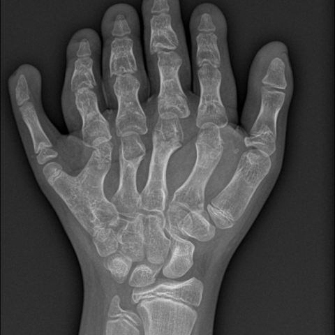
The radiography revealed a postaxial polydactyly with partial proximal synmetacarpalism V-VI. The 6th digit had only two phalanges (hypertrophic proximal and a distal). There was a fifth distal carpal bone (FDCB) at the ulnar side, articulating with the synmetacarpal. There was a right-left symmetry.
MRI moreover showed the more dorsal position of the distal 5th metacarpal. The tendon of the m. extensor digiti minimi (EDM) was running distally to the 5th finger, as the superficial and deep long flexor tendons. Although the hypothenar muscles were more centred around the 6th metacarpal, the tendons of the m. flexor (FDM) and m. opponens digiti minimi (ODM) were located at the 5th finger. Only the tendon of the m. abductor digiti minimi (AbDM) ran to the 6th finger. No large flexor tendons ran to the 6th finger.
Discussion
The Ellis-van Creveld Syndrome (EvC) (chondroectodermal dysplasia, six-fingered dwarfism) is a very rare genetic disorder of the skeletal dysplasia type, classified as a type of mesomelic limb shortening in the group of the short rib-polydactyly dysplasias (OMIM #225500). The reported incidence is in 1/1.500.000 births with an equal sex ratio. EvC is caused by loss-of-function mutations in the genes EVC or EVC2, the phenotype associated with the mutations in these genes being indistinguishable. The inheritance pattern is autosomal recessive.
Bilateral postaxial polydactyly is a constant finding. There are two types, a type A (well-formed digit) and a type B (rudimentary skin tag, designated as vestigial digit). Five subtypes are identified in type A [1]. One or more accessory carpal bones may be noticed, mostly on the ulnar side of the hamate bone (the so-called FDCB) [2, 3]. The FDCB is a remnant of more primitive tetrapod mammals [2]. The abnormalities tend to be symmetrical [3].
MRI showed, as well as plain radiography but without the use of ionizing radiation, a postaxial hexadactyly [4]. As usual, there was a partial segmentation of the 5th and 6th metacarpal and the extra digit had only two phalanges (hypertrophic proximal and distal). When the FDCB—as in our case—is not fused with the hamate bone, it articulates with the bases of the 5th and/or 6th metacarpals distally. MRI moreover showed that the EDM tendon was running distally adjacent to the 5th finger, as did the superficial and deep long flexor tendons. The tendons of the FDM and ODM ran adjacent to the 5th finger, whereas the tendon of the AbDM was located adjacent to the 6th finger. Also the more dorsal position of the 5th metacarpal is demonstrated clearly. Axial T1-SE and coronal T1-SE and T2-FS sequences are mandatory. In patients experiencing painful motion, more sensitive STIR sequences may be added - only for diagnostic reasons - in order to demonstrate dysfunctional bone marrow oedema due to the defective bony configuration. The longer examination time of MRI (versus plain radiography) poses no problem for the normal intelligent EVC patient.
When reconstruction of a type A is considered (not functional digit or for cosmetic reasons) MRI may precise pre-operative anatomy to plan the section planes and allow maximal preservation of the present muscle(s).
As far as we could establish, no previous MR studies of the hand or wrist in EVC have been published.
Differential Diagnosis List
Final Diagnosis
MR findings of postaxial polydactyly type A in EvC
Liscense
Figures
Plain radiography of the right hand

Plain radiography of the left hand
MRI of the right hand. Coronal SE T1-WI.

MRI (level marked on figure 3). Axial SE T1-WI.

MRI (level marked on figure 3). Axial SE T1-WI

MRI (level marked on figure 3). Axial SE T1-WI.

MRI at metacarpophalangeal level. Axial SE T1-WI.

Medical Imaging Analysis Report
I. Imaging Findings
The patient is an 11-year-old male previously diagnosed with Ellis-van Creveld syndrome (EvC), accompanied by postaxial polydactyly. The imaging examinations this time included a wrist X-ray and MRI.
- X-ray Radiography: Shows an additional sixth digit phalanx on the ulnar (little finger) side, indicating postaxial hexadactyly. Partial bony segmentation is observed between the 5th and 6th metacarpals, and the extra digit contains only proximal and distal phalanges. The positions of the scaphoid, lunate, triquetrum, pisiform, and hamate appear generally normal, but an extra carpal bone (FDCB) is visible on the ulnar side of the hamate, connecting with the bases of the 5th and/or 6th metacarpals.
- MRI: Sagittal and coronal sequences confirm the anatomical structures described above. The 5th metacarpal is relatively more dorsal in position, and in this case the additional carpal bone (FDCB) is not fused with the hamate but rather forms a separate articular surface. Regarding the flexor and extensor tendons, the extensor tendon follows the original 5th digit, while the flexor tendon shows a relative bifurcation between the 5th and 6th digits; the abductor digiti minimi (AbDM) tendon is positioned closer to the 6th digit. In addition, no obvious bone marrow edema or soft tissue swelling is seen on MRI.
- Fracture Evaluation: A subsequent assessment of a radial fracture shows no significant displacement or redisplacement, and the fracture line remains stable.
II. Potential Diagnoses
Based on the patient’s known diagnosis and imaging findings, possible or differential diagnoses include:
- Ellis-van Creveld syndrome (EvC) with postaxial polydactyly: The patient’s medical history, typical postaxial polydactyly, additional carpal bone (FDCB), and metacarpal deformities align with the characteristic features of EvC.
- Other types of short-rib polydactyly skeletal dysplasia: For example, Jeune syndrome, but such patients often show more distinctive thoracic and other skeletal abnormalities, which do not fully match this case’s X-ray findings or clinical history.
- Simple postaxial polydactyly: If there are no other EvC findings or systemic involvement, postaxial polydactyly alone may present. However, since the patient has a confirmed EvC background, it remains an auxiliary consideration.
III. Final Diagnosis
Considering the patient’s age, previously confirmed diagnosis of Ellis-van Creveld syndrome, typical postaxial polydactyly, and the multi-joint and tendinous structural changes indicated by X-ray and MRI, the most likely diagnosis is:
Ellis-van Creveld syndrome (EvC) with postaxial polydactyly (Type A).
IV. Treatment Plan and Rehabilitation
-
Treatment Strategy:
- If there is no functional impairment or cosmetic concern, follow-up observation can be performed. When the extra digit does not cause pain, restricted joint movements, or progressive deformity, surgery is not necessarily required.
- If the extra digit affects hand function or if there is a cosmetic need, surgical reconstruction or excision may be considered during adolescence or at an appropriate time, aiming to preserve or restore key tendon and ligament functions. MRI can assist in accurately locating tendons, nerves, and blood vessels before surgery.
- For concurrent radial fractures without noticeable misalignment, a cast or brace can be applied, with regular follow-up to monitor fracture healing.
-
Rehabilitation and Exercise Prescription:
- Goal: Maintain or improve joint range of motion and preserve muscle strength and coordination within the bounds of fracture healing and hand function.
- Gradual Progression (FITT-VP) Principle:
- Frequency: 1–2 sessions per day initially, possibly increasing to 2–3 sessions later, depending on individual needs.
- Intensity: Begin with low-resistance exercises, such as squeezing a simple handgrip ball or soft stress ball. If no significant discomfort or pain arises, gradually increase the intensity.
- Time: Each exercise session should last about 10–15 minutes initially, and can be extended to 20–30 minutes based on tolerance.
- Type: Include finger flexion and extension exercises, grip training, small-range wrist flexion and extension movements, and forearm pronation/supination exercises. If possible, a handgrip device or resistance bands may be used under the guidance of a professional rehabilitation therapist.
- Progression: As the fracture heals and function improves, reassess weekly or every two weeks. Gradually increase exercise intensity within safe limits, avoiding pain or risk of reinjury.
- Volume and Pattern: Adjust according to individual recovery, avoiding excessive fatigue and unstable joint loading.
- Special Considerations: EvC patients often have skeletal developmental abnormalities in other parts of the body, so regular monitoring of cardiopulmonary function and skeletal status is necessary. During fracture recovery, avoid significant impact or falls to reduce the risk of secondary injury.
Disclaimer:
The above report is a reference analysis based on current imaging and clinical information and cannot replace in-person consultation or professional medical advice. Specific diagnosis, treatment, and rehabilitation plans should be made after a thorough evaluation of the patient’s actual condition and professional assessments.
Human Doctor Final Diagnosis
MR findings of postaxial polydactyly type A in EvC