Osteoblastoma of the tibia that debuts as juxta-cortical osteolytic tumour


Clinical History
A 56-year-old male with a 1-year history of left leg pain that wakes him up at night. Pain has become constant over the last month.
No trauma was referred. No other relevant symptoms.
Imaging Findings
An x-ray, MRI and CT-guided biopsy were performed.
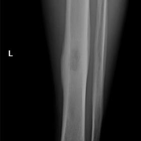
The frontal x-ray showed an intraosseous osteolytic lesion in the left tibia (type 1b geographic patron) with a thick continuous periosteal reaction (Figure 1a). The lateral projection showed the true location of the lesion sitting in the posterior cortex of the tibial diaphysis without medullary extension (Figure 1b) and with a narrow transition zone, indicative of a non-aggressive nature.
In the MRI, the lesion demonstrated no soft tissue mass. The bone findings consisted of the juxta-cortical lesion with peripheral oedema surrounding the bone marrow and soft tissues around the tibia (Figures 2a and 2b).
CT showed a cortical-based lesion with a solid periosteal reaction. The CT-guided biopsy confirmed the diagnosis of osteoblastoma (Figure 3A and 3B).
Discussion
Osteoblastomas are bone-forming tumours that account for 3% of all benign bone tumours [1]. Predominantly, affects young adults in the second decade of life, although it can appear at any age, with men’s predilection [2].
In the past, osteoblastomas and osteoid osteomas were classified as variants of a single tumour. Nowadays, these are considered two tumours that differ in location, clinical and radiological presentation and potential for progression [3].
Osteoblastoma is commonly located in the axial bones, whereas osteoid osteoma arises in the long bones. Osteoblastoma typically manifests as gradual, persistent pain that often does not alleviate with nonsteroidal anti-inflammatory drugs (NSAIDs), whereas the nocturnal pain characteristic of osteoid osteoma commonly shows significant improvement with NSAIDs [3].
Lesion size is a discriminating criterion, with osteoid osteoma being less than 1.5–2 cm in diameter and osteoblastoma having the potential to become larger (> 2 cm). In x-ray, osteoblastoma may have a bubbly appearance and internal calcification. It can associate soft tissue mass, cortical destruction and secondary aneurismal bone cyst-like changes in 20% [2,3]. CT can add matrix-related information, and although MRI may give some nonspecific information and tend to overestimate the lesion, it can be useful in describing bone and soft tissue oedema [4].
Based on the juxta-cortical location observed on radiography, differential diagnoses of metastasis, intracortical abscess, and benign primary tumour were entertained. While osseous metastases are notably more prevalent than primary osseous tumours, no prior history of malignancy was documented in this case. Furthermore, metastases typically manifest as multiple lesions, primarily affecting the axial or proximal appendicular skeleton. Additional signs include pathological fractures, soft tissue components and a broad transition zone suggestive of aggressive growth [5].
The subperiosteal abscess is a rare complication of bone infection and is more frequent in epiphyseal and subchondral locations secondary to intra-articular infection extending to the bone [6].
The continuous periosteal reaction and the area of central radiolucency with a narrow transition zone oriented the diagnosis towards a non-aggressive lesion. The diameter greater than 2 cm, the oval morphology and the oedema of the medullary bone and adjacent soft tissues suggested the diagnosis of osteoblastoma, which was confirmed after biopsy.
The patient was treated surgically with en bloc resection and remains asymptomatic 3 years later.
The most effective treatment is surgery. Preoperative embolisation is an adjuvant therapy but not chemotherapy or radiotherapy [7,8]. The risk of recurrence is from 15% to 25%.
Differential Diagnosis List
Final Diagnosis
Osteoblastoma
Liscense
This work is licensed under a Creative Commons Attribution-NonCommercial-ShareAlike 4.0 International License.
Figures
Radiograph of the left tibia and fibula


Axial PD-SPAIR MR


Axial non-enhanced CT


1. Radiological Findings
Based on the provided X-ray, CT, and MRI images of the left leg, a relatively regular oval lesion can be seen in the near-cortical region of the tibial shaft, measuring over 2 cm in diameter. On the X-ray, there is a mild cortical expansion and a continuous cortical response at the local site, with a relatively radiolucent area within the lesion and a well-defined boundary. CT imaging further reveals calcification or increased density within the bone, with limited destruction or thinning of the surrounding cortex. MRI shows abnormal signal changes in the lesion area, along with significant edema in the surrounding bone marrow and soft tissues, suggesting a more pronounced inflammatory or reactive change.
There is no obvious indication of a pathological fracture. Some secondary changes can be observed in the soft tissues, but there is no formation of a large soft tissue mass. Overall imaging features suggest a benign lesion: clear boundaries, localized periosteal reaction, and a lack of extensive infiltrative destruction.
2. Potential Diagnoses
- Osteoblastoma: Typically presents as a benign osteogenic tumor, which can affect the diaphysis of long bones. It often causes persistent pain that is not significantly relieved by NSAIDs. The lesion usually measures more than 2 cm in diameter. Imaging often shows lytic or “bubbly” changes, and the surrounding bone marrow and soft tissues frequently exhibit edema.
- Metastasis: Usually seen in patients with a history of malignancy. The lesions can be multiple and often involve the trunk or proximal long bones. In this case, the patient has no known history of malignancy. A solitary lesion with a well-defined boundary is less suggestive of metastatic disease.
- Subperiosteal Abscess: Rare, often related to infectious causes and may present with marked purulent manifestations. However, the patient’s blood tests and clinical symptoms do not indicate signs of acute or chronic infection. The lesion also does not show the aggressive destruction or significant fluid accumulation that would be expected in an abscess.
- Osteoid Osteoma: Similar to osteoblastoma but generally smaller than 1.5–2 cm, and it typically causes night pain that is significantly relieved by NSAIDs. In this case, the lesion is larger than 2 cm and the pain is not markedly responsive to analgesics, making this diagnosis less likely.
3. Final Diagnosis
Considering the patient is a 56-year-old male with persistent pain in the left leg, occasional worsening at night, and the imaging findings (an ovoid lesion larger than 2 cm, localized periosteal reaction, and notable soft tissue edema, yet with clear boundaries and confirmed by pathological biopsy), the most likely diagnosis is osteoblastoma. Pathological examination has confirmed this diagnosis, and the patient has recovered well following surgical resection.
4. Treatment Plan and Rehabilitation
Treatment Strategy:
- Surgical Resection: For osteoblastoma, especially those that are symptomatic or large, the best treatment option is complete curettage or en bloc resection (as performed in this case).
- Adjunctive Measures: If the lesion has a rich blood supply, preoperative embolization can be considered to reduce intraoperative bleeding. Chemotherapy or radiotherapy is not routinely recommended.
- Follow-Up and Imaging Review: Osteoblastoma has a certain recurrence rate (15%–25%), so regular imaging follow-up is required, especially within the first 1–2 years post-surgery.
Rehabilitation and Exercise Prescription:
Postoperative rehabilitation should follow a gradual and individualized approach, based on the patient’s soft tissue healing and bone healing status. The following is a general guideline:
- Early Stage (0–4 weeks post-surgery): Focus on protecting the affected limb and managing pain. Use assistive devices (e.g., crutches) to maintain partial weight-bearing. Encourage non-weight-bearing or light weight-bearing joint mobility exercises (e.g., ankle pumps, isometric quadriceps contractions).
- Mid Stage (4–8 weeks post-surgery): Once imaging confirms bone healing, begin to increase weight-bearing activities gradually. Emphasize walking training and simple lower limb strengthening (e.g., straight leg raises, slow squats). Focus on improving quadriceps and calf muscle strength, while controlling training intensity and frequency.
- Late Stage (8–12 weeks post-surgery and beyond): Gradually restore running, jumping, and daily activity capabilities. Increase weight-bearing and activity intensity step by step, such as extending walking time or introducing light jogging. For those with good overall fitness, advanced exercises (e.g., single-leg balance, resistance band exercises) may be introduced after adequate healing time.
- Injury Prevention and Safety: Rehabilitation should be guided by medical professionals or physical therapists. Protective braces or equipment should be used as needed. If pain recurs or swelling develops, return to the clinic promptly for evaluation.
This exercise prescription follows the FITT-VP principles: progressively adjusting Frequency, Intensity, Time, Type, and Progression based on the patient’s recovery status.
5. Disclaimer
The above report is a reference-based medical analysis derived from the current imaging and provided data. It is not intended to replace a face-to-face clinical diagnosis or professional medical opinion. In the event of any questions or changes in condition, please promptly seek care at a qualified medical institution for further evaluation.
Human Doctor Final Diagnosis
Osteoblastoma