

A 67-year-old man was referred to the orthopaedic department for further evaluation of osteoblastic bone lesions incidentally found on pelvis radiograph. Recent examinations revealed benign prostate hyperplasia. 99mTc bone scan (Fig. 6) showed positive radionuclide uptake, indicating possible osteoblastic metastases. His only complaint was moderate pain in the right knee.
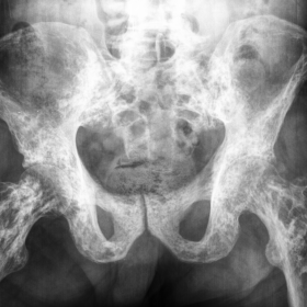
Plane radiography of the pelvis (Fig. 1) revealed the 3rd. grade osteoarthritis of hip joints according to Kellgren-Lowrence (KL) criteria associated with multiple, scattered, juxta-articular and metaphyseal, ovoid and longitudinal radiopacities.
Knee radiographs (Fig. 2) revealed KL grade 2 and KL grade 3 osteoarthritis of the left and the right knee. Ovoid sclerotic bone islands adjacent to inner cortices were detected in metaphyseal regions of femoral bones.
Shoulder joint radiographs (Fig. 3) detected the same sclerotic bone lesions in peri-articular and metaphyseal region of proximal humeral bones, clavicles and scapulae.
Control MDCT (Fig. 4) and MRI (Fig. 5) of the pelvis were performed to differentiate these lesions between osteoblastic metastases and benign bone dysplasias and to rule out prostate proliferation. The same ovoid and longitudinal symmetric sclerotic bone formations were detected in the pelvis and the proximal femurs.
Classic imaging findings of osteopoikilosis were associated with normal clinical, laboratory and histology results of benign prostate hyperplasia, excluding osteoblastic bone metastases.
Osteopoikilosis represents an autosomal dominant bone dysplasia that mimics osteoblastic bone metastases, and may lead to misdiagnosis and unnecessary treatment. Its incidence is 1 in 50, 000; it is seen in up to 6 per 100, 000 radiographs. [1] A mutation in gene LEMD3 at position 12q13 is responsible for this benign bone condition. [2]
Patients are usually asymptomatic or may complain of nonspecific articular pain in 15-20% of cases. Mostly, these lesions are discovered incidentally on radiographic imaging. [3] Radiographic findings of osteopoikilosis include the appearance of numerous symmetric, ovoid sclerotic foci, ranging from 2 to 10 mm, clustered in periarticular regions of tubular bones. The metaphyseal lesions may be longitudinal and located eccentrically, abutting the endosteal cortical surface. The main distribution of lesions in the pelvis is found about the acetabulum and mostly related to the orientation of the trabecular bone. The skull, mandible, clavicles and thoracic cage are spared. [4] Macroscopically, foci of compact bone within the cancellous bone are found. Histologically, the sclerotic nodular or star-like foci correspond to old and inactive condensation of cancellous bone with peripheral scant osteocytes. Osteoblasts and osteoclasts are in the central core of irregular trabeculae. [5]
Osteopoikilosis could be associated with skeletal and pea-sized fibrous skin changes as seen in Buschke-Ollendorf syndrome. It has been associated with scleroderma, syndactyly, dwarfism, mental retardation, and tuberous sclerosis. [1, 2] Mixed sclerosing bone dysplasia represents another condition, whereas triads of benign bone disorders: osteopoikilosis, osteopathia striata, and melorheostosis occur concomitantly.
The differential diagnosis includes conditions with radiographic appearance similar to those of
osteopoikilosis such as osteoblastic metastases, melorheostosis, osteopathia striata, mastocytosis, tuberous sclerosis and Paget disease. [4, 5] However, these bone conditions have additional distinctive physical findings.
Bone scans are mostly normal in osteopoikilosis, which has been used to differentiate it from metastatic bone disease. [6] There are a few previous reports of abnormal scintigraphy findings in osteopoikilosis, mimicking osteoblastic bone metastases. [7] Bone scan of a patient with bone metastastases usually reveals an increased uptake, with asymmetrical distribution when multiple sites throughout the skeleton are involved. The main problem is its low specificity of osteoblastic activity. Abnormal bone scan does not exclude the diagnosis of osteopoikilosis if the radiographic findings are characteristic of that entity.
Take Home Message:
Benign bone dysplasia that mimics osteoblastic bone metastases.
Usually asymptomatic.
Radiographic findings are characteristic and diagnostic.
Bone strength is normal.
Abnormal bone scan does not exclude the diagnosis of osteopoikilosis.
Osteopoikilosis (osteopathy condensans disseminata)
This work is licensed under a Creative Commons Attribution-NonCommercial-ShareAlike 4.0 International License.


Based on the provided pelvic, knee, and chest X-rays, as well as other imaging (CT, MRI, and 99mTc bone scans), the following features can be observed:
Considering the patient’s age, imaging characteristics, and medical history, the following differential diagnoses can be considered:
Taking into account the patient’s clinical presentation (only moderate pain in the right knee), along with the symmetrical, scattered distribution of small sclerotic foci on imaging, the absence of diminished bone strength, and only mild increases in uptake on the bone scan, it is still necessary to rule out malignant changes in the prostate clinically. However, the most likely diagnosis, based on the overall assessment, is:
Osteopoikilosis
If laboratory or pathological studies (e.g., checking for significantly elevated prostate-specific antigen or a bone biopsy) exclude any malignant findings, this diagnosis can be further confirmed.
Osteopoikilosis generally requires no special treatment since the patient’s bone strength is normal and the prognosis is favorable. In cases where imaging findings are incidental and no significant symptoms exist, a conservative follow-up approach is typically adopted. For this patient, the following recommendations are proposed:
This report is a reference analysis based on the currently available imaging and clinical information. It does not replace in-person consultations and specialists’ opinions. Always consult an appropriate specialist and combine their recommendations with the patient’s specific condition before making any treatment decisions.
Osteopoikilosis (osteopathy condensans disseminata)