Fused form of os intermetatarseum


Clinical History
A 37-year-old female patient was admitted to the orthopaedic clinic with persistent polyarthralgia (ankle and elbow), leg and ankle oedema and underwent foot X-ray, where a bone formation between proximal portion of first and second metatarsus was observed (Figures 1-3). Followed up at rheumatology consultation, the patient also presented temporomandibular joint arthralgia.
Imaging Findings
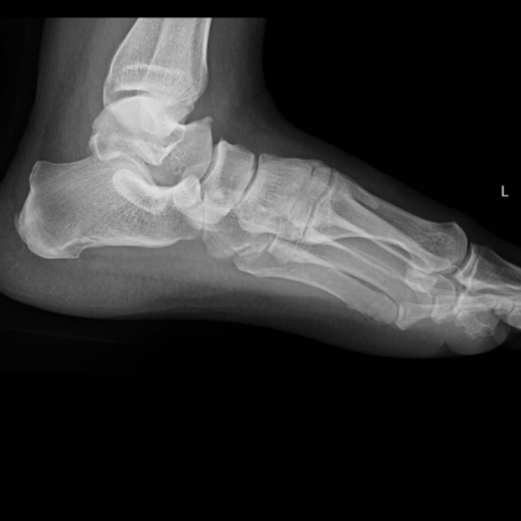
Besides degenerative alterations, namely in talus and navicular bones, a bone formation between proximal portions of the first and second metatarsus was observed on plain radiographs (AP, lateral and oblique projections) (Figure 1-3). This bone formation seems to be in relation with the second metatarsal base on dorsomedial surface. Foot CT (Figures 4-6) confirms the presence of this bone formation as an apparent exostosis process in the dependence of the dorsomedial surface of second metatarsal base with 30mm of long axis. This bone formation presented characteristics of an incomplete metatarsus, including shape, orientation and presence of narrow bone inside of the cortical bone, compatible with os intermetatarseum in fused form with the second metatarsal base. A ligament tissue was observed between the distal intermetatarseum part and first phalanx.
Discrete hallux valgus (a complication of this condition [1]) signals were present, namely a sclerotic metatarsophalangeal articular surface and discrete phalanx deviation, and only the intermetatarsal angle measurement was superior to normal.
Discussion
Os intermetatarseum is a rare accessory bone of the foot, usually found between first and second metatarsal bones [1, 2, 3]. According to several authors the incidence range is 1.2 to 14% [3]. This disparate range of incidence probably is related with heritability, what make this variant more frequent in some populations [1].
Commonly, os intermetatarseum are asymptomatic, but rare cases had been reported as painful, once they caused anterior tarsal tunnel syndrome in relation with overuse or trauma of the dorsal part of the foot [1, 4, 5]. Therefore, mostly they are diagnosed accidentally, as it happened in this case.
The os intermetatarseum can present various forms: free-standing type, a completely independent ossicle with no articular connections with neighbour bones; articulating type, presents a synovial joint with first metatarsal, second metatarsal, first cuneiform or even with two or three of these bones; or fused form, the rarest type of os intermetatarseum, where the accessory metatarsal is fused with first metatarsal base, second metatarsal base or first cuneiform [2].
In first instance, plain X-ray projections are used to find and describe. On the other hand, CT scan gives a better description of os intermetatarseum. Therefore, those are useful tools in the diagnosis of this normal variant. Os intermetatarseum shows up in the image as a bone formation, located commonly between the first and the second metatarsal. The relation with neighbour structures depends on the type.
The fused form of os intermetatarseum, although rare, is the most easily recognizable form because it tend to present long osseous projections [2]. On plain radiographs it could be observed as an apparent exostosis between first and second metatarsal in dependence of the first or second metatarsal base, or even the first cuneiform. In this patient it was observed in dependence of the second metatarsal bone (Figures 1-3). Due to a high overlap of structures in the metatarsal bases, plain radiographs could not distinguish the fused form of other forms of os intermetatarseum. In CT scan, the imaging findings are the same, but we could better describe the relation between neighbouring structures, and also describe with precision the type of os intermetatarseum present, as it is possible to observe in Figures 4 to 7. CT scan also contributes to rule out false positives. MRI, even not performed in this patient, could have an important role in the study of the ligament tissue (tendon-like form) between distal part of the os intermetatarseum and first proximal phalanx.
Differential Diagnosis List
Final Diagnosis
Os intermetatarseum (fused form)
Liscense
This work is licensed under a Creative Commons Attribution-NonCommercial-ShareAlike 4.0 International License.
Figures
Foot AP X-ray projection

Foot oblique X-ray projection

Foot lateral X-ray projection
CT axial images

CT sagittal images

CT coronal images

3D volume rendering

Medical Imaging Analysis Report
1. Imaging Findings
Left foot X-ray images (Figures 1–3) and CT scans (Figures 4–7) of the patient show a bony structure with a clear boundary between the proximal parts of the first and second metatarsals, resembling an extra bony prominence. It is closely associated with the base of the second metatarsal, and 3D CT reconstruction suggests a possible fusion of this bone with the base of the second metatarsal. No obvious swelling of surrounding soft tissue or signs of bone destruction are noted.
Apart from this special bony structure in the foot, the articular surfaces are generally intact, with no obvious fractures or significant bony abnormalities. The patient also has multiple joint pain (ankle and elbow joints) and ankle swelling, but there is no clearly identified source of pain from the local foot imaging.
2. Potential Diagnoses
- Os intermetatarseum: Based on the imaging characteristics, an additional bony structure between the first and second metatarsals is most consistent with Os intermetatarseum, a rare accessory bone of the foot, especially its fused type.
- Bony spur or osteophyte at the base of the metatarsal: Abnormal proliferation or bony spur commonly occurs in chronic overuse or degenerative changes. However, the fused appearance in this case looks well-defined and not like a typical degenerative spur.
- Benign bony tumor (e.g., ossifying fibroma, osteochondroma): Usually there is a more distinct pattern of bone destruction or proliferative change with noticeable mass effect. In this case, the imaging findings lean more toward a congenital variant rather than a tumor.
3. Final Diagnosis
Combining the patient’s age, clinical symptoms, and imaging findings, the possibility of a bony tumor or proliferative osteophyte can be ruled out. The morphology and location of this extra bone strongly suggest a fused Os intermetatarseum. This conclusion aligns with the fact that the patient has multiple joint pain in other areas without a corresponding significant pathological lesion in the foot, indicating this bony variant is more likely an incidental radiological finding.
4. Treatment Plan and Rehabilitation
For Os intermetatarseum that does not cause significant symptoms, no special treatment is typically needed. If pain or discomfort in the dorsal or plantar region of the foot occurs, consider the following treatment and rehabilitation plans:
- Conservative Treatment
- Use arch supports or orthotic insoles to reduce local pressure.
- Oral or topical non-steroidal anti-inflammatory drugs (NSAIDs) to relieve symptoms.
- Hot compresses or local physical therapy to help reduce soft tissue tension.
- Surgical Intervention
- If persistent pain or nerve impingement (e.g., involvement of the dorsal plantar fascia or forefoot) is caused by the accessory bone or its position and conservative treatment is ineffective, consider surgical removal or correction.
- The surgical approach mainly involves resection of the extra bone and decompression, usually with good prognosis.
- Rehabilitation and Exercise Prescription
- Initially, focus on range-of-motion exercises for the joints (e.g., dorsiflexion and plantarflexion of the ankle), performed 2–3 times a day for 5–10 minutes each session.
- After inflammation and pain subside, gradually add foot and lower limb strengthening exercises, such as standing on tiptoes or alternating toe-heel stands, 2–3 times per week for 10–15 minutes each time.
- If permitted by the patient’s overall condition, slowly resume low-impact aerobic exercises (e.g., swimming, stationary cycling) 2–3 times per week for 20–30 minutes each session, adjusting the intensity appropriately.
- As muscle strength and endurance improve, the exercise intensity or load can be gradually increased in accordance with the FITT-VP principle (Frequency, Intensity, Time, Type, Volume, Progression), under the guidance of a professional.
- For other joint symptoms (ankle, elbow), a comprehensive management approach should be considered to avoid overuse and further joint pain or injury.
5. Disclaimer
This report provides a preliminary analysis based on existing medical imaging and clinical information. It is for reference only and cannot replace an in-person consultation or professional medical opinion. If symptoms persist or worsen, promptly visit a qualified medical institution and follow specialized medical advice for further evaluation and treatment.
Human Doctor Final Diagnosis
Os intermetatarseum (fused form)