


An 18-year-old female dancer complained of recurrent right dorsal foot pain and stiffness, without history of trauma. Clinical examination revealed limited subtalar mobility.
Since the plain radiographs of the right foot (not available to view) were read as normal, the patient underwent magnetic resonance imaging (MRI) to define the cause of the symptoms.
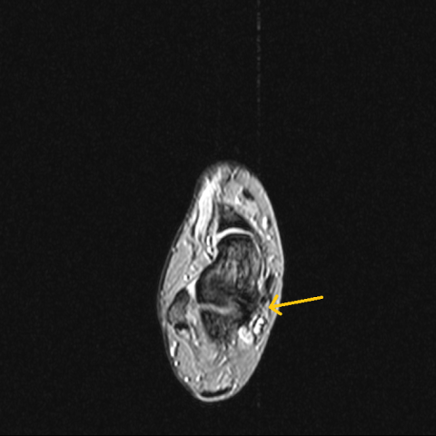
MRI of the right foot revealed a talocalcaneal bridge that included the middle facet of the subtalar joint with continuity of the marrow signal intensity, indicating an osseous talocalcaneal coalition (Fig. 1, 2, 3). Short-inversion-time inversion recovery (STIR) images showed bone marrow oedema that involved the talocalcaneal bridge and the opposing surfaces of talus, calcaneus and medial malleolus (Fig. 3).
Talocalcaneal coalition represents together with calcaneonavicular coalition the most common type of tarsal coalition, which is an abnormal fusion of two or more tarsal bones and has a prevalence in the general population of about 1% [1, 2].
This disease may have a congenital origin, consisting in failure of complete segmentation in the early stages of primitive mesenchyme with lack of normal joint formation [3].
Talocalcaneal coalition may be osseous, cartilaginous or fibrous and it usually involves the middle facet of the subtalar joint, at the level of the substentaculum tali [1]. Many authors report that a progressive osseous bridging occurs with increasing age, the reason why various histologic components may coexist [4].
A recent study introduces a new type of talocalcaneal coalition, in which an os sustentaculum is part of the extra-articular bridge between talus and calcaneus [5].
Talocalcaneal coalition is usually asymptomatic in early childhood; tarsal pain and stiffness appear during adolescence, with the increase in ossification, and are usually associated with physical activity [3]. A talocalcaneal coalition with os substentaculum may be suspected in all patients with an os substentaculum who complain of pain of the medial talocalcaneal joint area [5].
Imaging plays an important role in the detection of talocalcaneal coalition. The radiographic signs are: talar beak, narrowing of the posterior subtalar joint, rounding of the lateral talar process and “C sign”, seen on lateral radiographs, consisting of a C-shaped line that represents the bridge between talar dome and substentaculum tali [2]. Talocalcaneal coalitions may be difficult to detect on plain radiographs, so its diagnosis and characterization require computed tomography (CT) or MRI [4].
CT, offering multiplanar capability, is useful to define the size, location, and orientation of talocalcaneal coalitions and is able to identify an os substentaculum through the osseous bridge [4, 5]. However, CT is not recommended in young patients, because of the use of ionizing radiation.
MRI, using T1-weighted, fast spin-echo proton density–weighted, and fast spin-echo T2-weighted images, allows us to detect talocalcaneal coalition and to define its type: cartilaginous, fibrous and bony, with or without os substentaculum. Moreover, thanks to fluid-sensitive sequences, it’s easily possible to identify bone marrow oedema, which could be associated to talocalcaneal coalition [4, 5].
In cases with non-disabling symptoms the management of talocalcaneal coalition may be conservative, with orthoses, physical therapies and anti-inflammatory medications. If all conservative treatments fail, surgery may be indicated [3].
In our case the patient was conservatively treated, although her dancing career had to end too early.
Talocalcaneal coalition
This work is licensed under a Creative Commons Attribution-NonCommercial-ShareAlike 4.0 International License.


Based on the patient’s MRI scans (multiple sequences including T1-weighted, PD-weighted, and T2-weighted images), the following main features can be observed:
Combining the patient’s symptoms (recurrent dorsal foot pain, stiffness, no obvious history of acute trauma) and imaging findings, the following diagnoses or differential diagnoses can be considered:
Considering the patient’s age (18-year-old dancer), recurrent foot pain and stiffness, limited subtalar joint mobility on clinical examination, and MRI findings of an abnormal bridge between the talus and calcaneus along with minimal adjacent bone marrow edema, the most likely diagnosis is:
Talocalcaneal coalition at the middle facet of the subtalar joint, possibly involving an “os sustentaculum.”
To further clarify the proportions of bony and cartilaginous tissue, a CT scan can provide precise visualization of the bony bridge. However, in younger patients, MRI is often preferred initially to avoid or reduce radiation exposure.
For talocalcaneal coalition, management should be personalized based on symptom severity and the patient’s functional needs.
Suggestions for Rehabilitation and Exercise Prescription:
Disclaimer:
This report is based on current clinical and imaging information for reference in medical knowledge and does not substitute for an in-person consultation or professional medical diagnosis and treatment. If you have any concerns, please consult a specialist or seek further medical evaluation promptly.
Talocalcaneal coalition