


17-year-old Thai boxer athlete presented with pain on the lateral foot edge. Pain was elicited by palpation of the fifth metatarsal and also by plantar flexion and resisted eversion. Plain radiographs and magnetic resonance imaging were performed.
Plain radiographs revealed absence of union of the secondary ossification centre of the base of the fifth metatarsal (Fig. 1). Rest, ice and analgesia was prescribed.
Despite conservative treatment, he continued to experience lateral foot pain six months after. A complementary magnetic resonance imaging (MRI) was performed. Sagittal fat-suppressed T2-weighted images (WI) showed bone marrow oedema at the secondary ossification centre and the adjacent metaphysis of the fifth metatarsal (Fig. 2). Axial T1-WI showed slight widening of the growth plate between the secondary ossification centre and the base of the fifth metatarsal (Fig. 3). Based on the imaging findings, the diagnosis of Iselin’s disease was made.
Iselin’s disease previously known as traction apophysitis of the secondary ossification centre of the fifth metatarsal is an osteochondrosis of the foot. Dr. Iselin described this disorder in 1912. [1] It is thought to result from repetitive traction of the peroneus brevis tendon and the fifth metatarsal base. The term apophysitis is a misnomer because it is a non-inflammatory process. The pathogenesis consists of disruption of the vascular supply of the fifth metatarsal apophysis, resulting in avascular necrosis, with bone resorption, followed by healing and recalcification. [2]
Growing athletes are at increased risk of developing this type of injury, especially when they do sports with running, jumping or repetitive inversion of the forefoot. The roundhouse kick is a famous Thai boxer kick, in which the hips are rotated, followed by pivoting of the supporting non-kicking foot. This creates repetitive stress on the fifth metatarsal of the non-kicking foot.
Clinically there is tenderness at palpation over the insertion of the peroneus brevis tendon with no ecchymosis. Resisted eversion and plantar flexion are painful. [3]
The apophysis has an inferolateral location and is best viewed on oblique radiographs. The secondary ossification centre is seen as a bony fleck oriented longitudinal to the long axis of the fifth metatarsal. [2]
In case of Iselin’s disease, widening of the growth plate may be seen as well as fragmentation of the apophysis. MRI is the most sensitive imaging modality, as it demonstrates bone marrow oedema before radiographic changes are visible. Bone marrow oedema within the apophysis and the metaphysis of the fifth metatarsal base are suggestive for the diagnosis. In addition, widening of the growth plate and associated soft tissue oedema may be observed.
The differential diagnosis consists of Jones fracture, avulsion fracture and os vesalianum. Jones fracture is transversely oriented and more distally at the meta-diaphyseal junction. An avulsion fracture is the acute equivalent of Iselin’s disease, with avulsion of the apophysis at the insertion of the peroneus brevis tendon. Os vesalianum is a well-corticated accessory bone, and is more proximally located [4].
Iselin’s disease is managed conservatively, and responds well to NSAIDs, ice and resting.
To the best of our knowledge this is the first case of Iselin’s disease reported in a Thai boxer. MRI is more sensitive than plain radiographs for early detection of Iselin’s disease, which may allow early treatment and may prevent long-term complications such as non-union and subsequent pain.
Iselin’s disease of the fifth metatarsal
This work is licensed under a Creative Commons Attribution-NonCommercial-ShareAlike 4.0 International License.
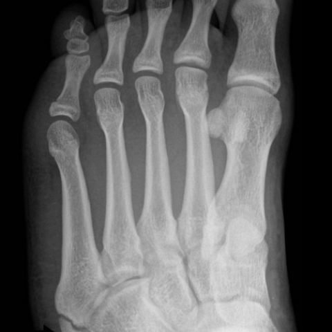


From the provided foot X-ray images (anteroposterior and oblique views), an abnormal shape and position of the secondary ossification center (apophysis) at the lateral edge of the base of the fifth metatarsal can be observed. There is widening and fragmentation of the apophyseal line, but no obvious fresh fracture line is seen. Soft tissue swelling is not significant.
MRI shows abnormal bone marrow signals in and around the apophyseal region of the fifth metatarsal base: on T2-weighted or fat-suppressed sequences, this presents as high signal intensity, suggesting local bone marrow edema. There is also extra-articular soft tissue edema around the apophyseal line. These findings correspond to local injuries caused by repeated pulling or flexion-extension stresses.
Based on the patient’s training characteristics (Muay Thai with high-frequency rotational and weight-bearing foot movements), the location of symptoms (lateral edge of the fifth metatarsal base), and the imaging features, the following are the main potential or differential diagnoses:
Combining the patient’s age (adolescent), the nature of the sport (frequent kicking and support movements in Muay Thai), clinical presentation (tenderness along the lateral foot border, pain with resisted eversion and plantar flexion), and imaging findings (apophyseal fragmentation, bone marrow and adjacent soft tissue edema), the most likely diagnosis is: Iselin disease (traction apophysitis of the fifth metatarsal base).
Treatment Strategy:
1. Conservative Treatment: Most cases of Iselin disease can be managed with rest, avoidance of aggravating exercises, local ice massage, and NSAIDs to relieve symptoms. If necessary, short-term use of a foot brace or soft support device can reduce lateral traction on the foot.
2. Physical Therapy: After acute pain subsides, perform ankle and foot muscle balance training, stretching of the fibular muscles (especially the peroneus brevis), and low-load resistance exercises to help reduce pulling stress on the fifth metatarsal base.
3. Surgical Intervention: Rarely, in cases of severe injury or recurrent chronic pain affecting athletic performance, surgery may be considered, although it is generally uncommon.
Rehabilitation/Exercise Prescription Recommendations (FITT-VP Principle):
• Frequency (F): During the acute phase, reduce weight-bearing and avoid painful movements. One session per day of light foot activity or simple foot and ankle stretching is recommended. After symptom relief, gradually increase to moderate strength and stability training 3–4 times per week.
• Intensity (I): Start with mild intensity (bodyweight support, no pain) and slowly increase the load as symptoms improve. Avoid high-impact or sustained eversion movements.
• Time (T): Initially 5–10 minutes per session focused on ankle flexibility and foot muscle strengthening, gradually extending to 20–30 minutes depending on patient tolerance and pain levels.
• Type (T): Include core foot muscle training (e.g., toe gripping, light resistance for foot inversion and eversion), balance training (single-leg stance or using a balance board), as well as low-impact aerobic exercises like swimming or cycling.
• Progression (P): Increase load or refine techniques (e.g., adding more balance challenges or resistance) only when the patient can perform the current exercises without significant pain or discomfort. If pain worsens, pause and adjust the plan.
• Volume and Progress (V & P): Monitor weekly symptom changes and functional improvements. Schedule regular follow-ups to evaluate apophyseal healing and foot function, and steadily increase training volume and complexity.
This report is based solely on existing information for reference and cannot replace an in-person consultation or the professional diagnosis and treatment advice of a physician. If you have any questions or if symptoms worsen, it is recommended to seek medical attention promptly and undergo further examination.
Iselin’s disease of the fifth metatarsal