

A 41-year-old man from Pakistan with a 2-year history of a steadily growing lump in the region of the second finger of his left foot. No drug allergies. No toxic habits. Hepatitis C, received treatment with peg IFN for 12 months. Denied other medical and surgical history of interest.
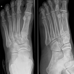
Plain radiographs showed translucence at interphalangeal joint and moderate sclerosis of the second finger of the left foot. X-ray imaging shows classically a so-called "snow-melt" form.
Ultrasound images of the left foot showed a mainly hypointense lesion containing small, discrete, hyperechoic foci.
MRI revealed a soft tissue mass in the subcutaneous plane rounding the second finger of the left foot with a deformity of the interphalangeal joint of the 2nd finger showing irregularly joint surface, deformity and bone destruction. The mass showed conglomerate areas of multiple, discrete, small 2-5 mm round hyperintense lesions, which were separated by a low-signal-intensity rim in the subcutaneous plane of the second finger of the left foot. Within many of these hyperintense lesions, there was a central low-signal-intensity dot. A separate focus of involvement was seen in the medial aspect of the foot.
¨Mycetoma is a debilitating chronic granulomatous infection by true fungi (eumycetoma) or by bacteria of the order Actinomycetales (actinomycetoma). It affects the subcutaneous tissues and may extend to involve deeper structures¨ [1].
Madurella mycetomi in Asia and Streptomyces somaliensis or pelletieri in Sudan have been also described as aetiological agents in the literature. Actinomycetoma due to Actinomadura
madurae is an extremely rare condition in temperate zones [2].
It is prevalent in the tropical and subtropical regions. The infection painlessly burrows deeply until it reaches the bone.
Early diagnosis is sometimes difficult. If untreated, it can lead to significant destruction and deformity. Biopsy or microbiological culture can provide the diagnosis, but this may not always be possible.
Mycetoma is characterized by the formation of aggregates of the organism, known as ¨grains¨, which are found within abscesses surrounded by abundant granulation tissue.
¨Ultrasound showed distinct hyperechoic foci within a hypoechoic mass¨. [3].
This could account for the appearance we observed on MRI of conglomerates of small (2-5 mm) round hyperintense lesions, representing the granulation tissue, surrounded by low-signal-intensity rim, representing intervening fibrous septa. The central low-signal-intensity dot is the result of susceptibility effect caused by the presence of fungal grains. ¨This is a unique appearance, that appears to be highly suggestive of mycetoma and it is called ¨dot-in-circle¨ [4].
Some authors have proposed ¨to classify the pattern, extent and severity of bone involvement in mycetoma of the foot. In this classification, stage 0 indicates the presence of soft-tissue swelling without bone involvement. Stage I refers to the extrinsic pressure effects on the intact bones in the vicinity of an expanding granuloma. Stage II results from irritation of the bone surface without actual intraosseous invasion. Cortical erosion and central cavitation occur in stage III. If the disease spreads longitudinally along a single ray, stage IV is established; horizontal spread along a single row represents stage V. Multidirectional spread due to uncontrolled infection is classified as stage VI.¨ [5]
Our case would correspond to a stage II.
In the mycetoma involving the soft tissues of the foot, antimicrobial therapy is sometimes curative. When there is bone involvement, non-surgical cure is unlikely and partial resection or amputation may be required.
Early diagnosis of this disease and bone involvement is therefore essential for appropriate management.
Our patient was treated with streptomycin and dapsone. Eight months later, the patient no longer needed analgesics, and there was a clear clinical, radiological, and laboratory improvement. He continues with chemotherapy for at least 18 months.
Madura foot with dot-in-circle sign.
This work is licensed under a Creative Commons Attribution-NonCommercial-ShareAlike 4.0 International License.


Based on the provided foot X-ray, ultrasound, and MRI images, the following characteristics can be observed:
Considering the patient’s clinical presentation (a slowly enlarging mass on the second toe), endemic area, and the imaging findings, the main differential diagnoses include:
After considering the clinical course, the characteristic “dot-in-circle” appearance on imaging, clinical examination results, and the epidemiological context of the region, the most likely diagnosis is:
Mycetoma (“Madura foot”), most likely Actinomycetoma caused by actinomycetes.
Further microbiological workup, including culture, histopathological examination, or molecular testing, can help confirm the specific pathogen and guide targeted therapy.
Standard treatment for mycetoma involves long-term combined antimicrobial or antifungal therapy to control the infection, and surgical intervention may be considered if necessary:
This report is intended for reference only and cannot replace in-person consultation or tailored professional advice and prescriptions by a physician. If any new symptoms appear or the condition changes, please seek specialist advice or visit a hospital promptly.
Madura foot with dot-in-circle sign.