Calcinosis in limited cutaneous systemic sclerosis


Clinical History
A 62-year-old woman with a 10-year history of limited cutaneous systemic sclerosis presented with insidious pain and swelling of the left thumb’s fingertip. Physical examination revealed sclerodactyly and a tender nodule on the distal phalanx of the first finger.
Imaging Findings
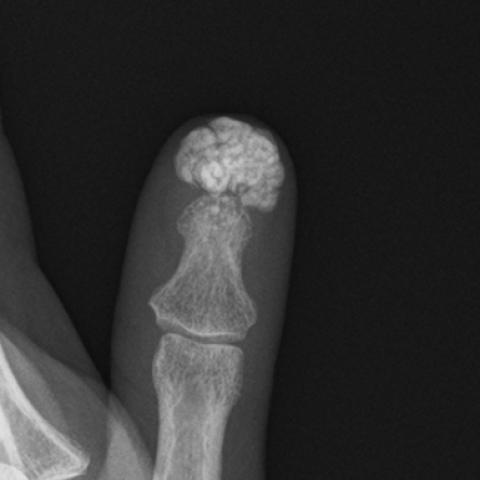
Radiographic imaging of the left hand (Figures 1a and 1b) and thumb (Figure 2) revealed a well-defined calcification in the soft tissue overlying the distal phalanx of the left first finger, measuring approximately 1.5 cm. Periarticular osteopenia was also observed, particularly evident at the thumb (Figure 1b). Cortical thinning and blurring of the margins were noted in the proximal interphalangeal joint of the ring finger (Figure 1a). No other significant findings were identified.
Discussion
Background
Systemic sclerosis (SSc) is an immune-mediated disorder characterised by skin and connective tissue fibrosis and vascular dysfunction [1–2]. Calcinosis is a common manifestation in limited SSc and often develops years after the initial diagnosis of SSc [2–4]. Its pathophysiology is unclear, but digital vasculopathy appears to contribute to its development [2,3].
Clinical Perspective
Calcinosis may significantly affect patients’ quality of life due to pain, skin ulceration and potential infection [3]. Early detection is crucial for timely decision-making on optimal management [2–3].
Imaging Perspective
Radiography is the preferred diagnostic tool for confirming calcinosis, as it efficiently identifies calcified deposits in soft tissues [3–4].
Outcome
Treatment of calcinosis in SSc remains challenging [2–3]. Therapeutic options include symptomatic management and preventive measures against ulceration and infection [3]. In severe cases, surgical excision is considered [2–3].
Teaching Points
- Radiographic imaging allows the identification of calcinosis in systemic sclerosis (SSc).
- Calcinosis is a common manifestation of SSc and can impact the quality of life.
- Management of calcinosis remains challenging, with treatment primarily focused on symptom relief and prevention of complications.
Differential Diagnosis List
Final Diagnosis
Calcinosis (in the setting of limited cutaneous systemic sclerosis)
Liscense
This work is licensed under a Creative Commons Attribution-NonCommercial-ShareAlike 4.0 International License.
Figures
Left-hand radiograph


Left first finger radiograph
Medical Analysis Report
1. Radiological Findings
1. From the provided anteroposterior (AP) and lateral X-rays of the left thumb, a high-density shadow, appearing as a mass-like calcification with relatively clear boundaries, is visible near the nail bed area of the distal phalanx.
2. The adjacent distal phalanx shows adequate bony structure without notable destruction or fracture lines. The bone shape is not evidently deformed, but mild thickening of the subcutaneous soft tissue is observed, suggesting possible chronic inflammation or localized fibrosis.
3. No obvious pathological changes are noted in the other phalanges, joint spaces, or adjacent tissues.
2. Potential Diagnoses
- Calcinosis cutis related to systemic sclerosis (scleroderma)
- The patient has a longstanding, 10-year history of limited cutaneous systemic sclerosis (lcSSc), which predisposes to digital ischemia and microcirculation impairment, leading to subcutaneous deposition of calcium salts.
- The X-ray finding of a localized, dense calcific mass within soft tissue aligns with common features seen in calcinosis cutis.
- Gouty tophus
- Gouty tophi can present as pinpoint or mass-like calcifications; however, these are typically associated with a history of gout and elevated serum uric acid levels.
- This case lacks supporting metabolic or gout history, and the X-ray presentation is more consistent with calcinosis cutis.
- Other soft tissue calcifications
- Such as ectopic calcifications due to repeated trauma, local infection, or scar tissue, although these usually do not occur in the context of long-term systemic sclerosis.
3. Most Likely Final Diagnosis
Taking into account the patient’s age, sex, extended history of lcSSc, and radiographic findings, the most likely diagnosis is: Calcinosis cutis associated with systemic sclerosis.
4. Treatment Plan and Rehabilitation
1. Treatment Strategy
- Conservative Treatment: For mild or localized symptoms, focus on symptomatic management including anti-inflammatory pain medication and local care to prevent skin breakdown and secondary infection.
- Pharmacological Therapy: In cases with significant vasospasm (Raynaud’s phenomenon), vasodilators or other medications that improve microcirculation may be considered under the guidance of a rheumatologist; calcium channel blockers might also be used.
- Surgical Intervention: If the calcified mass is large, repeatedly infected, or severely impairs hand function, surgical excision may be evaluated.
- Regular Follow-up: Monitor changes in the lesions and soft tissues to detect new calcifications or complications in a timely manner.
2. Rehabilitation and Exercise Prescription (FITT-VP Principles)
- Frequency: 2-3 times per week, focusing on exercises that enhance finger flexibility and grip strength.
- Intensity: Begin with low intensity and gradually increase according to pain and swelling levels; avoid heavy loads or intense movements.
- Time: 15-20 minutes per session, possibly divided into segments; incorporate active finger joint movement and flexion-extension exercises into daily routines.
- Type:
- After warm water immersion or heat application, perform active and passive stretching of the finger joints to alleviate skin tightness caused by scleroderma and to prevent joint contractures.
- Use a grip ball or soft objects for squeezing exercises to improve thumb and small hand muscle strength and fine motor coordination.
- Progression: Gradually prolong the exercise duration or introduce mild resistance equipment (e.g., resistance bands, soft balls with varying resistance levels) as symptoms improve.
- Volume: Maintain an appropriate amount of exercise and closely monitor hand fatigue or pain. If there is notable redness, swelling, or discomfort, reduce intensity and seek professional evaluation.
Note: Because systemic sclerosis often involves vascular and skin fragility, pay attention to skin temperature and digital circulation, and avoid excessive stimulation or trauma.
5. Disclaimer
This report provides a reference analysis based on the provided medical history and imaging data and cannot replace in-person consultation or professional medical guidance. If you have any concerns or if symptoms worsen, please seek medical attention or consult a specialist promptly.
Human Doctor Final Diagnosis
Calcinosis (in the setting of limited cutaneous systemic sclerosis)