

The patient was a 46-year-old woman with history of rectal cancer stage IV diagnosed in 2013, treated with an anterior rectal resection followed by adjuvant chemo and radiotherapy. She had undergone multiple chemotherapy treatments to the present date. The disease had metastasized to her lungs but no other sites of metastasis were documented.
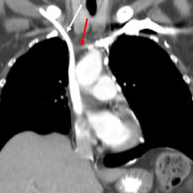
Thoracic CT after IV contrast showed an obstruction of both innominate veins and the initial segment of superior vena cava, just above the confluence with azygos vein (figure 1), without evidence of local adenopathies or any extrinsic mass. We interpreted these findings as fibrosis related obstruction due to the multiple previous catheterizations of chemotherapy treatments.
Not surprisingly, we also observed exuberant collateral circulation owing to the referred venous obstruction, involving common and less frequent pathways: azygos - hemiazygos system, internal mammary - inferior epigastric vessels, vertebral venous plexuses and the pericardiophrenic vein (draining to inferior vena cava).
The most interesting finding was a triangular shaped uptake centered at basivertebral foramen of the first dorsal vertebrae, without any notorious atenuation changes at unenhanced scan of the same level (figures 2 and 3).
Superior vena cava syndrome (SVCS) is a clinical entity caused by an obstruction of venous flow coming from the head, upper extremities and upper thorax at the level of the superior vena cava (SVC) or of both innominate veins.
The clinical presentation depends on the velocity of installation, meaning that acute occlusions often lead to more severe symptoms, in contrast to slow developing obstructions which are usually milder at presentation [1].
The classic symptoms are head, neck and upper limbs swelling. Dyspnoea may also be a common complaint, consequently to larynx and pharynx oedema [2].
In slow evolving obstructions, there is enough time to build venous collaterals which divert blood away from the occluded SVC. Various venous systems are involved in this process. By order of importance, the azygos-hemiazygos system, internal mammary to inferior epigastric veins, lateral thoracic to superficial epigastric veins and the vertebral venous plexuses are the usual pathways implicated [3, 4]. On rare occasions, collaterals can drain to the pulmonary system, functioning as a right-to-left shunt, which has important clinical implications by raising the risk of stroke and high cardiac output state [5]. Cyanosis can be evident in those cases.
Nowadays, the majority of SCVS cases are malignant-related, especially as a consequence of direct compression from upper lung tumours with mediastinal invasion [6]. Other malignancies associated with SVCS include anterior mediastinal tumours, mediastinal metastases (especially from breast cancer), lymphoma with mediastinal involvement and in scarce cases, primary tumours of the vena cava [7].
Infectious diseases were an important cause of SVCS, either by mediastinal inflammation and fibrosis, such as in histoplasmosis and tuberculosis, or by vascular injuries like in syphilitic aortic aneurysms [7]. The first description of superior vena cava syndrome was made by a Scottish anatomist, William Hunter, who in 1757, reported a syphilitic aneurysm of the ascending aorta compressing the neighbor SVC [8]. With the advent of antibiotics this paradigm changed, and the remainder of benign aetiologies are more commonly due to medical interventions, such as placement of central venous lines and pacemakers, which predisposes to SVC thrombosis and fibrosis [7].
Pseudo-bone metastasis (vertebral plexus vascular congestion)
This work is licensed under a Creative Commons Attribution-NonCommercial-ShareAlike 4.0 International License.


According to the provided chest CT images, the following observations can be made:
In summary, the images indicate compression or obstruction of the SVC, as well as possible bone metastatic changes in part of the spine.
Considering the patient’s known history of stage IV rectal cancer, pulmonary metastases, and current imaging findings, the following differential diagnoses are suggested:
Given the patient’s advanced (stage IV) rectal cancer, existing history of pulmonary metastasis, and the imaging findings indicating SVC compression or narrowing, mediastinal mass/enlarged lymph nodes, and vertebral involvement, the most likely diagnosis is:
Superior Vena Cava Syndrome (SVCS) caused by malignant (metastatic) tumor, accompanied by spinal bone metastases.
If necessary for further clarification of the tumor’s histopathological nature or to exclude other causes (such as a newly developed tumor or certain lymphoma), mediastinal lymph node or suspicious lesion biopsy and bone lesion sampling should be considered.
As the patient has advanced cancer, a safe and feasible rehabilitation plan should be developed according to their actual condition. Special attention should be paid to the following:
This report is based solely on the current imaging and clinical history provided for reference. It does not replace an in-person consultation or the final judgment of a professional physician. A specific treatment plan should be formulated and implemented by a qualified medical team, taking into account the patient’s clinical presentation, laboratory results, and any additional examinations.
Pseudo-bone metastasis (vertebral plexus vascular congestion)