Pachydermoperiostosis - A rare radiologic case


Clinical History
The patient had complaints of clubbing, swelling of multiple joints and thickening of the facial skin since adolescence. The patient complained of excessive sweating and feeling of heat and burning sensation in palms and soles. Thyroid profile, growth hormone assay, tests for syphilis and smears of skin for AFB were unremarkable.
Imaging Findings
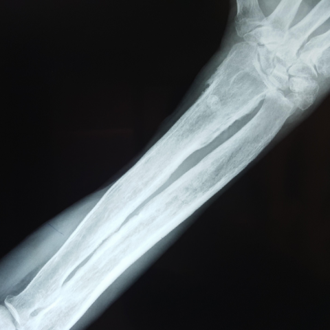
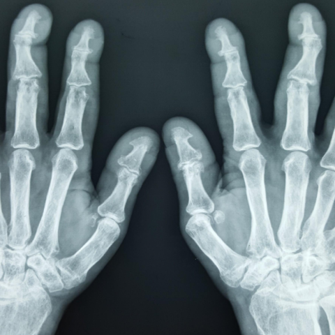
Symmetric, exuberant, shaggy subperiosteal bone formation was seen in both forearms and legs. There was expansion of the distal ends of radius and ulna and the proximal ends of tibia and fibula, with a reduction in the radiocarpal and femorotibial joint spaces. There was reduction in joint spaces of the proximal and distal interphalangeal joints. Periarticular osteopenia and resorption of the distal phalanges was noted with associated soft tissue swelling of distal fingers and toes. Widening of the base of distal phalanx of all fingers of both hands was seen. Enlargement and remodelling of sesamoid bones in both hands and feet was noted.
The images showed collapse of calcaneum and calcification of tendoachilles. There was a symmetric and shaggy periosteal reaction along the lower lateral aspect of the iliac bones.
There was evidence of metaphyseal widening of the bilateral femur with cortical thickening and widening of the shafts.
There was mild hyperostosis of the skull bones in the parietooccipital region. The sella turcica appeared normal.
Discussion
PDP is a rare form of hypertrophic osteoarthropathy with no known cause and hence it is called idiopathic or primary hypertrophic osteoarthropathy (PHO). PDP accounts for 3–5% of cases of hypertrophic arthropathy and affects males more often than females (7:1).
In up to one-third of the patients, PDP occurs as an autosomal dominant hereditary disease. Some case reports suggest that it may be an X-linked disease. [1]
Deficiency of the prostaglandin transporter (SLCO2A1) has been characterized as the main cause of PHO.
Touraine et al. described three forms of PDP, viz, classic or complete form, with skin and skeletal changes; incomplete form, with skeletal changes but no dermal findings; and forme fruste with dermal changes but no skeletal findings. [2]
Facial involvement occurs in the form of thickening of the facial skin and scalp, with prominent folds on forehead and cheeks. Sometimes, the scalp takes on an undulating appearance and shows prominent grooves, the appearance called cutis verticis gyrata. Cutis verticis gyrata can also be seen in a variety of other conditions, including neurofibromatosis, DM, myxoedema, acromegaly, etc., and in syndromes including Turner's syndrome, tuberous sclerosis, etc., hence it is not pathognomonic for PDP. [3]
Skeletal findings include symmetric, shaggy subperiosteal bone formation in the long bones, especially of forearm and leg. Involvement of the epiphyseal region distinguishes it from the secondary form, in which epiphyses are usually spared. [4] There is widening of the ends of bones, especially at wrist and knee joints. A prominent feature is enlargement of distal part of the digits with resorption of distal phalanges and calcification of ligaments and interosseous membranes. In later stages, cortical thickening with narrowing of medullary cavity may be seen. Enlargement of sinuses may be seen uncommonly. Bone scintigraphy may reveal increased tracer uptake by the cortex in diaphyseal and metaphyseal regions. [5, 6] Hyperostosis of the calvaria and skull base bones is common. [4]
Joints affected in PDP show swelling due to joint effusion, with evidence of chronic nonsuppurative inflammation. There is reduction in joint spaces, with relative preservation of articular surfaces. Late-onset deformities may occur, especially in the digits. [5]
Spinal manifestations are unusual but have been described.
Variants of PDP include Rosenfeld-Kloepfer syndrome, Currarino idiopathic osteoarthropathy and a localized form with only the radiographic features of PDP in the lower extremities. [6]
Treatment is limited to NSAID, steroid, or colchicine therapy to alleviate arthralgias and retinoids for dermal changes. Surgical treatment is limited to plastic surgery for cosmetic indications. [5, 6, 7].
Differential Diagnosis List
Final Diagnosis
Pachydermoperiostosis
Liscense
This work is licensed under a Creative Commons Attribution-NonCommercial-ShareAlike 4.0 International License.
Figures
AP radiograph of both knee joints

AP radiograph of forearm
AP radiograph of both hands
Lateral radiograph of ankle joint

Radiograph of pelvis with both hips

Lateral radiograph of skull

AP radiograph of foot

USG examination of knee

1. Imaging Findings
Based on the provided multi-site X-ray images (including bilateral knees, forearms, hands, ankles, pelvis, skull, etc.), the following main features are observed:
• Thickening of long bone cortical shafts and periosteal reactions: Noticeable, symmetrical periosteal proliferation and cortical thickening are seen in the forearm (radius and ulna) and lower leg (tibia and fibula).
• Enlargement of distal phalanges and soft tissue thickening: Bilateral hand X-rays show terminal expansion of the phalanges and possible marked soft tissue thickening, suggesting clubbing.
• Relatively preserved joint spaces: Most joint spaces do not show significant narrowing. The articular surfaces appear generally intact, with no obvious erosive changes.
• Thickening of the skull bones: There may be varying degrees of hyperostosis in the skull base and skullcap, as evidenced by thickening on skull X-ray. If accompanied by scalp folding, clinical findings may suggest “cutis verticis gyrata.”
• Pelvis and ankle joints: The pelvic X-ray also shows some degree of cortical thickening or periosteal reaction; ankle and foot X-rays indicate distal bone thickening, and hypertrophy of terminal digits.
Overall, these findings are consistent with a systemic, symmetrical periosteal proliferation. Combined with clinically observed thickening of the skin, clubbing, and joint swelling, this suggests a characteristic hypertrophic osteoarthropathy.
2. Potential Diagnoses
Based on the patient’s clinical symptoms (clubbing, joint swelling, facial skin thickening since puberty) and the above imaging findings, the main differential diagnoses include:
1) Primary Hypertrophic Osteoarthropathy (Pachydermoperiostosis, PDP/PHO): Also known as idiopathic hypertrophic osteoarthropathy. Classic features include skin thickening (pachydermia), periosteal proliferation (periostosis), and clubbing. Imaging often reveals notable proliferative changes in the diaphyses and metaphyses of long bones, frequently involving the epiphyses, without obvious cardiopulmonary pathology as the cause.
2) Secondary Hypertrophic Osteoarthropathy: Commonly associated with thoracic tumors, chronic pulmonary or cardiac diseases. Periostosis is often concentrated around the metaphyses, but other primary disease features may also be present. If previous examinations (e.g., chest screening) were unremarkable, a secondary cause is less likely.
3) Acromegaly: Usually associated with elevated growth hormone, leading to joint and soft tissue thickening and characteristic facial changes. However, laboratory tests (GH, IGF-1, etc.) are typically abnormal in acromegaly. Since this patient’s growth hormone and thyroid function tests are normal, this is less likely.
4) Other Endocrine/Metabolic Causes: Such as myxedema (hypothyroidism) or other rare disorders. Typically, clinical presentation and laboratory indices would show corresponding abnormalities.
3. Final Diagnosis
Taking into account the patient’s long-term (since puberty) progressively developing skin thickening, clubbing, and joint swelling, in the absence of thyroid dysfunction, acromegaly, or secondary causes (e.g., pulmonary or cardiac diseases), along with classic findings of generalized periosteal proliferation and joint changes, the most likely diagnosis is “Primary Hypertrophic Osteoarthropathy (Pachydermoperiostosis)”.
For further confirmation, genetic testing (e.g., identification of SLCO2A1 mutations) or additional clinical biochemical and imaging follow-up could be considered.
4. Treatment Plan and Rehabilitation
Treatment Strategy:
• Medication for symptomatic relief: Primarily non-steroidal anti-inflammatory drugs (NSAIDs), corticosteroids, or colchicine to alleviate joint pain, swelling, and inflammation. For patients with pronounced skin thickening, retinoids may be considered.
• Surgical and cosmetic interventions: For patients with significant facial or scalp folds and aesthetic concerns, evaluation for plastic surgery may be appropriate.
• Health education and follow-up: Emphasize skin care, monitor joint function changes, and perform routine assessments of bone density and joint status.
Rehabilitation and Exercise Prescription:
Considering the patient’s joint swelling and bone thickening, exercises should focus on joint protection, preserving range of motion, and maintaining muscle strength. Specific recommendations are as follows:
1) Initial Stage:
• Frequency: 3–4 times per week;
• Intensity: Low to moderate intensity (e.g., basic joint mobility exercises, low-load resistance training, 20–30 minutes of walking), including gentle range of motion exercises;
• Duration: Each session lasts 20–30 minutes, depending on joint tolerance;
• Mode: Choose low-impact aerobic exercises such as swimming, using an elliptical trainer, or supported walking, combined with core and upper/lower limb strength training.
2) Progressive Stage:
• Frequency: Gradually increase to 5 times per week;
• Intensity: Increase resistance or speed appropriately, without triggering significant joint pain;
• Duration: Up to 30–45 minutes per session;
• Mode: Continue low-impact aerobic exercise and add flexibility and strength training, such as resistance band exercises in seated or supine positions.
Throughout rehabilitation, it is essential to regularly assess joint pain and swelling. If there is significant discomfort or worsening deformities, patients should seek medical review and adjust exercise accordingly.
5. Disclaimer
This report is based on the available imaging and clinical information for preliminary analysis only. It is intended for academic and clinical reference and does not replace face-to-face consultation or professional medical advice. If the condition changes or you have further concerns, please seek medical attention for appropriate examinations and treatment.
Human Doctor Final Diagnosis
Pachydermoperiostosis