Otto pelvis


Clinical History
A 35-year-old female patient came with complaints of gradually increasing bilateral hip pain and restriction of motion since 7 years. No past history of trauma or steroid intake. Blood counts were normal.
Imaging Findings
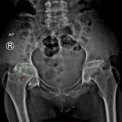
On X-ray PBH AP view, the distance between medially located acetabular line and laterally located ischio-femoral line was 7 mm on the right side and 3 mm on the left side. The centre edge angle of Wiberg was 70 degrees on the right side and 58 degrees on the left side, suggestive of right moderate and left mild protrusio acetabuli. (Figure 1)
There was severe symmetrical reduction in the joint space with medial migration of femoral heads, femoral head articular surface deformity in the form of mild collapse, flattening and irregularity, subchondral sclerosis and femoro-acetabular osteophytes (grade IV osteoarthritis). (Figure 2)
On MRI with T1WI, PDfatsat, STIR and T2WI, the above findings were confirmed. Additional findings noted were near complete denudation of articular cartilage, subchondral cysts, thin curvilinear hypo-intense subchondral lines in weight bearing portion of femoral epiphyses (possible subchondral fractures), patchy hyper-intensities (on STIR) involving bilateral femoral heads and juxta-articular acetabulum with minimal bilateral joint effusion.(Figures 3 and 4)
Discussion
Otto pelvis or arthrokatadysis is an unusual form of primary osteoarthritis with primary protrusio acetabuli, described first by German pathologist Otto in 1824 [1], frequently affecting young women. Familial factors like failure of ossification or remodelling of weak acetabulum are implicated in its aetiology. In one-third of patients it is bilateral [2].
Usually osteoarthritis is associated with supero-lateral subluxation of the femoral head. However, in the 20% of individuals who present with primary acetabular protrusion, there is concentric joint space loss and medial intra-pelvic displacement of the medial wall of the acetabulum and the femoral head with degenerative changes established at an early age [3]. Patients may be asymptomatic or may present with hip (or rarely knee) pain, restriction of movements and joint stiffness [4].
Plain AP radiographs of bilateral hip joints are adequate in diagnosis. MR arthrograms with radial sequences may be required pre-operatively for assessing cartilage injury, marrow changes and any associated features of pincer femoro-acetabular impingement [5].
Normally, on an AP radiograph, the medial wall of the acetabulum lies 2 mm lateral to the ilio-ischial line (Kohler’s line) in males and 1 mm medial to this line in females. If the medial wall of acetabulum protrudes medial to the ilio-ischial line by 3 mm in males or 6 mm in females, it favors the diagnosis of protrusion. Protrusion is graded as mild (1 to 5 mm), moderate (5 to 10 mm) and severe (10 to 15 mm) with reference to the ilio-ischial line. Center Edge angle of Wiberg is an angle formed by a line drawn from the centre of the femoral head to the outer edge of the femoral roof and a vertical line drawn through the centre of the femoral head. Protrusion is present if the CE angle is greater than 35 degrees. Normally, the CE angle is around 25 degrees while an angle less than 20 degrees suggests dysplasia [5].
In cases without cartilage degeneration, open surgical dislocation with osteochondroplasty of the acetabular rim and the femoral neck is recommended. Valgus inter-trochanteric osteotomy may be indicated in cases with inadequate femoroacetabular clearance. In cases with early cartilage degeneration on MRA, osteochondroplasty is not sufficient and osteotomy of the pelvis, femur, or both depends on the individual morphology. In adult patients with advanced degenerative changes, total hip arthroplasty is the treatment of choice [6].
Differential Diagnosis List
Final Diagnosis
Otto pelvis
Liscense
This work is licensed under a Creative Commons Attribution-NonCommercial-ShareAlike 4.0 International License.
Figures
1. Xray PBH AP view- Bilateral protrusio acetabuli
Xray PBH AP view- Grade IV osteoarthritis

T2WI coronal MRI of hip joint

T1WI STIR Axial MRI of hip joint

Medical Imaging Analysis Report
1. Imaging Findings
From the provided bilateral hip anteroposterior (AP) X-ray images and MRI scans, the following key features are observed:
1. Both hip joints show medial displacement of the acetabula. The femoral heads and the medial margins of the acetabula protrude significantly inward (“protrusio”), and the joint space between the acetabular roof and the femoral head is narrowed on the medial side.
2. According to the center-edge (CE) angle measurements, the CE angle in both hip joints exceeds 35° (e.g., up to 57°), which indicates excessive acetabular coverage of the femoral head.
3. Combining the MRI findings shows increased coverage at the acetabular rim, and some articular surfaces may show early signs of degeneration, requiring higher-resolution evaluation of the cartilage.
4. There is no obvious fracture or extensive bony destruction around the hip joints. The margins of the acetabulum and the femoral head articular surface may show mild degenerative changes.
2. Potential Diagnoses
Based on the patient’s 7-year history of gradually worsening bilateral hip pain and restricted range of motion, with no clear history of trauma or steroid use, along with laboratory tests showing no significant inflammation or metabolic abnormality, and considering the imaging findings, the following diagnoses or differential diagnoses should be considered:
- Primary protrusio acetabuli (Otto pelvis or primary acetabular protrusion): Caused by developmental dysplasia or defective bone remodeling of the acetabulum. It is commonly seen in young females and often presents with bilateral and symmetrical inward protrusion of the acetabula in the absence of overt external triggers.
- Secondary protrusio acetabuli: Conditions such as rheumatoid arthritis or metabolic bone diseases (e.g., achondroplasia) may lead to acetabular protrusion, but they are usually accompanied by other laboratory or systemic findings. In this case, neither the blood tests nor the patient history suggests these factors.
3. Final Diagnosis
Considering the patient’s sex, age, imaging characteristics (bilateral acetabular protrusion with markedly increased center-edge angles), and the absence of evident secondary causes, the most likely diagnosis is:
Primary protrusio acetabuli (Otto pelvis).
4. Treatment Plan and Rehabilitation Program
1. Treatment Strategy
- Surgical Intervention: Chosen based on the condition of the cartilage and severity of symptoms:
- If there is no significant cartilage degeneration, an open hip procedure can be considered, combined with acetabular rim recontouring (to address excessive acetabular coverage).
- If there is more severe femoroacetabular impingement or insufficient coverage of the femoral head, an osteotomy (e.g., intertrochanteric valgus osteotomy or pelvic osteotomy) may be performed after assessing the alignment to improve hip motion and reduce pain.
- For adult patients with marked degenerative changes, total hip arthroplasty (THA) can be considered to relieve pain and improve function.
- Conservative Treatment: When symptoms are not severe or during pre- and post-operative care, nonsteroidal anti-inflammatory drugs (NSAIDs) for pain relief and physical therapy (e.g., heat therapy, ultrasound, muscle strengthening) can be employed as appropriate.
2. Rehabilitation / Exercise Prescription Recommendations
The principles of rehabilitation and exercise training should follow an individualized and progressive approach (FITT-VP: Frequency, Intensity, Time, Type, Progression, and Individualization). For this case, possible considerations include:
- Initial Stage (Preoperative or Early Postoperative):
- Frequency: 3–4 times per week.
- Intensity: Low intensity, focusing on non-weight-bearing or partially weight-bearing exercises, such as seated or supine hip range-of-motion exercises. Avoid deep hip flexion.
- Time: 20–30 minutes per session, combined with heat packs or other supportive therapies.
- Type: Range-of-motion exercises (active and passive) and isometric contractions of the hip muscles (e.g., quadriceps, gluteal muscles, core stabilization).
- Intermediate Stage (Gradual Weight-Bearing and Strengthening):
- Within pain tolerance, increase hip range of motion and engage in aerobic activities (e.g., wall squats, seated cycling, swimming) to strengthen hip and core muscle groups.
- Frequency: 3–5 times per week.
- Intensity: Moderate intensity; ensure proper form and gradually increase duration or resistance.
- Time: 30–45 minutes per session.
- Late Stage (Functional Recovery and Return to Daily Activities):
- Based on hip function and strength recovery, progressively add standing exercises, light resistance band workouts, etc.
- Incorporate balance and coordination training, such as single-leg stance to improve proprioception.
- If pain or discomfort worsens, reduce exercise volume or suspend the relevant activities and seek medical advice.
Throughout the rehabilitation process, it is essential to monitor hip pain, signs of inflammation, and changes in lower limb alignment. If significant pain or a sudden decline in the range of motion occurs, seek prompt evaluation or adjust the treatment plan accordingly.
5. Disclaimer
This report is based on the provided clinical history and imaging findings, combined with reasonable medical reasoning and analysis, and is intended only as a clinical reference. The specific treatment plan requires an in-person consultation, further diagnostic evaluations (e.g., pathology or related laboratory data), and a comprehensive assessment by a qualified physician. If you have any questions, please consult a professional physician or visit a reputable medical institution.
Human Doctor Final Diagnosis
Otto pelvis