Case report of bilateral dysplasia epiphysealis hemimelica (DEH) of talus


Clinical History
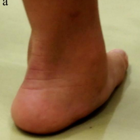
A 5-year and 4-month-old boy consulted our hospital with left leg pain, caused by torus fracture of the distal tibia. During the treatment, both his flat feet and a slight bony prominence in both medial sides of the talus were noticed. There was neither pain nor discomfort at his foot bottom.
Imaging Findings
Flatfoot and valgus deformity of calcaneal were found (Fig.1). Plane X-ray pictures revealed deformity of the talus and bony protrusions at the medial side of the talus with a slight punctate change as a mirror lesion in both tali (Fig 2, 3). His younger sister’s plain AP radiograph of both ankles showed almost the same bony protrusion formation in the medial side of both tali (Fig.4). Other relatives, including parents had no special notes. MRI revealed an iso-intense mass covered by cartilage arising from both the medial side of the talus neck and the head in T1-Weighted image and T2-Weighted image (Fig.5). Abnormal intensity, indicating suspicion of malignancy, was not detected. The talo-navicular joint was delineated between the deformed head of the talus and the navicular. No dislocation of the talo-navicular joint was found (Fig.6).
Discussion
Background
DEH was first reported by B. J. Mouchet A in 1926. In 1950, Trevor D. reported DEH using the term Tarso-epiphyseal aclasis. In 1956, Fairbank first used the term dysplastic epiphyseal hemimelica [1]. DEH is a rare developmental disorder of epiphyseal osteocartilaginous growth. Usually an osteocartilaginous mass arises from one side and on the medial side of the body. It most often presents at the knee and ankle; involvement of the upper extremities is rare. Aetiology and heredity are unknown. The incidence has been reported as one per million. The ratio between boys and girls is 3:1. DEH has the following three different types: local, classic and generalised types. In the current case, the local type would be applicable, but “bilateral local type” might be a more proper category. The word ''hemimelica'' is derived from two greek words: ''hemi'' meaning half and ''melos'' meaning limb. Nothing in the name would prevent applying it to bilateral lesions.
Clinical perspective
There are only 5 case reports we could find in the past, including one case of both tali lesions [2, 3, 4, 5]. Those locations, the presence of a mirror lesion, radiographic, MRI and pathological findings and comorbidity were shown in Table 1. By the place that a tumour produces, the symptoms are various. Sometimes the clinical symptom is deformity or stiffness of the ankle, knee, and so on. Sometimes it will be pain. In the present case, the only symptom is hump and flat feet deformity. There was no pain nor stiffness.
Imaging perspective
Regarding radiological characteristics, an irregular mass containing punctate ossificasion arising from the epiphysis is typically detected. During the infant or toddler age, it may be only an epiphyseal widening. At adolescent or adult age, an irregular bone mass, containing multi-centric radiodensity is seen. Gradually, the mass becomes adhesive to normal bone. In the current case, radiological findings were close to the ones typically found in the older age group, rather than in the younger age category. MRI showed iso-intensity bony mass covered with cartilage.
Outcome
Treatment of DEH varies depending on location, severity, symptoms, dysfunction, deformity and so on. If there are mild symptoms, deformities, or dysfunctions, conservative therapy will be chosen (like an arch support applied for flat foot in the current case). Operative treatment will be considered if there is persistent pain related to the deformity and/or dysfunction of the adjacent joint.
Differential Diagnosis List
Final Diagnosis
Bilateral dysplasia epiphysealis hemimelica
Liscense
This work is licensed under a Creative Commons Attribution-NonCommercial-ShareAlike 4.0 International License.
Figures
Plane AP radiograph

Plane AP radiograph of feet (axial view)

Younger sister’s plain AP radiograph

5 cases reported in the past and current study

Photographs from the posteromedial side

Plane MRI coronal view


Plane MRI sagittal view


1. Radiological Findings
Based on the bilateral ankle X-rays and MRI images provided, irregular bony protrusions or “mass-like” changes are observed in both tali (particularly medially). These protrusions are continuous with the normal bone structure, and multiple centers of calcification or increased bone density can be seen within the lesions. MRI indicates that the bony protrusions are covered by a cartilaginous-like signal and remain in continuity with the normal talus. Overall, the morphology resembles exostoses seen in osteochondroma, with bilateral and relatively symmetrical lesions. Clinically, the patient presents with flat feet and a slight palpable bump on the medial aspect of the ankle, but there is no obvious soft tissue swelling or localized soft tissue lesion. Additionally, mild wedge-shaped compression or buckling (a torus fracture-like change) is seen in the left distal tibia, consistent with a typical torus (buckle) fracture.
2. Potential Diagnoses
- Dysplasia Epiphysealis Hemimelica (DEH), also known as Trevor disease. This is a rare, epiphyseal chondroma-like lesion occurring in the pediatric population, often involving unilateral or medial portions of the epiphysis, resembling “osteochondroma.” Occasionally, contralateral “mirror” lesions can present. Clinical symptoms range from asymptomatic to mild joint deformities. Considering the bilateral, medial talar bony protrusions with a cartilaginous cap in this case, DEH is highly suspicious.
- Osteochondroma. Commonly seen in the metaphysis near the growth plate, presenting as an exostosis with a cartilage cap. However, occurrence in tarsal bones such as the talus is relatively rare, and it typically appears as a single or asymmetrical lesion. Bilateral and symmetrical involvement of the epiphyseal region is less common, making this diagnosis less likely here.
- Enchondroma. Usually occurs within the medullary cavity (intramedullary), presenting as cartilage-like densities or signals inside the marrow space, and is most frequently found in the phalanges of the hand or the long bones. In this case, the lesions are on the epiphyseal surface and show continuity with the cortex, hence do not favor enchondroma.
- Other rare epiphyseal cartilage disorders, such as atypical cartilage tumors or epiphyseal cartilage degeneration, might be considered. However, based on the patient’s age, bilateral location, and the specific imaging features, these are less plausible.
In summary, the most important diagnostic consideration focuses on an abnormality of the epiphyseal cartilage. Considering the clinical and imaging findings, Dysplasia Epiphysealis Hemimelica (DEH) is most consistent with these features.
3. Final Diagnosis
Taking into account the patient's age, clinical presentation (left distal tibia fracture, bilateral flat feet, and a mild medial ankle protrusion without significant pain or joint dysfunction), imaging findings (bilateral medial talar bony outgrowth covered by a cartilaginous cap), and literature descriptions of this condition, the most likely diagnosis is:
Dysplasia Epiphysealis Hemimelica (DEH)
If further differential diagnosis or evaluation is needed, additional pathological biopsy or periodic follow-up imaging can be considered to assess progression.
4. Treatment Plan and Rehabilitation Program
4.1 Treatment Strategy
- Conservative Treatment: Given the mild current symptoms presented by the patient, primarily flatfoot deformity and a painless bony prominence of the medial talus, a supportive intervention such as arch supports or orthotic insoles can help maintain foot arch stability and reduce extra stress on the affected area. Regular follow-up is recommended to observe skeletal development and lesion progression.
- Surgical Indications: If the patient develops persistent pain, significant deformity progression, or impaired joint function (e.g., ankle instability or reduced range of motion), surgical excision of the bony protrusion with realignment of the joint may be considered. The timing of surgery generally depends on the child’s growth and the severity of symptoms.
4.2 Rehabilitation and Exercise Prescription
During conservative management, it is recommended to follow a gradual, individualized rehabilitation program according to the FITT-VP principle:
- Type of Exercise: Emphasize low-impact, joint-stabilizing activities, such as swimming, cycling, or lower-limb range-of-motion exercises. Include exercises targeting core foot muscle groups (e.g., foot arch strengthening).
- Frequency: Approximately 3–5 sessions per week, increasing gradually according to the child’s tolerance.
- Intensity: Light to moderate intensity is advisable, avoiding excessive impact from running or jumping that could stress the ankle joint. Use orthotic supports or protective taping when necessary.
- Time: Each exercise session could last approximately 15–30 minutes, potentially divided into segments to prevent fatigue.
- Progression: Adjust the duration and difficulty of exercises based on follow-up assessments and the child’s subjective feedback. Monitor ankle stability and foot arch changes. If any significant pain or swelling occurs, reduce exercise volume and seek medical evaluation.
Additionally, parents and the patient should be vigilant about new lower limb injuries (e.g., sprains, fractures), and schedule regular follow-ups to ensure proper bone development and control of the lesion.
5. Disclaimer
The above content is for reference only and does not replace an in-person consultation or professional medical advice. If you have any questions or if symptoms change, please consult a medical professional or specialized healthcare facility promptly.
Human Doctor Final Diagnosis
Bilateral dysplasia epiphysealis hemimelica