Invasive vertebral haemangioma


Clinical History
Male patient, 44 years of age, with no medical history of interest. Consultation for chronic lumbociatalgia associated with paresthesias of both lower limbs.
Imaging Findings
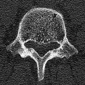
No significant findings are observed on the lateral radiograph of the lumbar spine. In the axial plane of the CT, the involvement of the entire vertebral soma of L5 is observed, with the presence of multiple punctate foci giving the impression of salt and pepper (Fig. 1). In the sagittal and coronal reconstructions, dense and lucid interspersed bands are observed, giving the impression of bars (Fig. 2a, b). In MRI, signal hyperintensity is observed in both the T1 and T2-weighted images, as well as in the fat-suppressed images (STIR), showing a lesion that occupies the entire vertebral body of L5, which breaks its posterior wall and invades the medullar canal by more than 50%, compressing the descending and emerging nerve roots at this level (Fig.3a-c).
Discussion
Vertebral haemangiomas are frequent lesions, which are observed in about 10% of the autopsies performed. Usually they are incidental findings and do not produce symptoms. They do not constitute a neoplasm, but rather a congenital anomaly derived from the sequestration of mesodermal embryonic tissue in the vertebrae [1, 2].
In rare cases (1% of all vertebral haemangiomas) they can cause symptoms, which is termed invasive hemangioma. They are characterised by bone expansion, extraosseous extension, local blood flow alterations and can be the origin of pathological fractures, associated with a deficit/neurological symptoms [1, 2]. The most affected vertebrae are the thoracic vertebrae. In the imaging studies, typical images characterised by radio-lucidity of the affected vertebral body will be observed on plain radiography; on CT, in the axial plane there is a dotted appearance of salt and pepper, and in the sagittal planes of cell bars. In MRI the signal will be given by the presence of fat and vascular tissue arranged between the trabeculae of the bone, and the intensity of the signal will depend on the proportion of the content of these. Invasive haemangiomas usually contain less fat and a greater vascular stroma [1, 2, 3]. With the contrast medium they tend to enhance in an avid way, which is why they can be confused with metastasis [4], however, with the presence of the other image features previously mentioned, the diagnosis can be reached, preventing patient's anguish and moreover unnecessary treatment.
The treatment can be surgical, especially in large lesions; with radiotherapy use; or through techniques performed by interventional radiologists such as sclerosis or embolisations of the vessels, or percutaneous vertebroplasty with injection of cement semiliquid that undergoes a solid state polymerisation process, which gives a mechanical support to the vertebra, improving pain and decreasing the risk of fractures [2, 3]. In the case of our patient, this last technique was performed with satisfactory results (Fig. 4).
Differential Diagnosis List
Final Diagnosis
Invasive vertebral haemangioma
Liscense
This work is licensed under a Creative Commons Attribution-NonCommercial-ShareAlike 4.0 International License.
Figures
CT

CT


MRI


Vertebroplasty

Medical Imaging Analysis Report
1. Imaging Findings
Based on the provided CT and MRI images, typical “salt and pepper” or “grid-like” density changes can be seen within the lower lumbar vertebrae (mainly involving the L4 or L5 vertebral bodies in the images). In the sagittal view, vertical stripe-like alterations are observed, and in certain areas, relatively thickened trabeculae are visible. On MRI sequences, both T1 and T2 signals within the vertebral body appear elevated, suggesting the coexistence of fatty components and vascular spaces. In addition, there is a mild expansile change with localized protrusion around the vertebral body, but no obvious vertebral compression fracture is noted. Considering the clinical symptoms (chronic low back and leg pain with bilateral lower limb sensory disturbances), these findings indicate that the vertebral lesion is associated with nerve irritation. Overall, the imaging features are highly characteristic of a “vertebral hemangioma.”
2. Differential Diagnoses
-
Vertebral Hemangioma
Reason: This type of lesion is often congenital or developmental in origin, characterized by an overgrowth of vascular tissue within the vertebral body. It is commonly found in the thoracic and lumbar spine. Typical imaging features include a “salt and pepper” or “polka-dot” appearance on CT, and high signal on MRI due to fatty and vascular components. If invasive, it may show vertebral expansion, soft tissue involvement, and neurological symptoms. -
Vertebral Metastasis
Reason: Frequently seen when malignant tumors spread to the vertebrae. Abnormal density or signal on CT or MRI is common, typically accompanied by significant bone destruction or a soft tissue mass. If there is prominent enhancement on contrast imaging, metastasis should be considered. However, metastatic lesions usually lack the distinct trabecular pattern characteristic of hemangiomas. -
Multiple Myeloma
Reason: A hematologic malignancy that can involve the vertebrae, often presenting with “punched-out” lesions. Compared to vertebral hemangioma, its CT and MRI findings differ considerably, usually lacking the classic “grid-like” or “salt and pepper” sign.
In summary, based on the imaging findings and the patient’s chronic low back pain combined with sciatica-like symptoms, the most likely diagnosis is vertebral hemangioma, potentially exhibiting local invasive features causing nerve involvement.
3. Final Diagnosis
Considering the patient’s age (44 years), chronic low back and leg pain with bilateral lower limb sensory abnormalities, and the typical imaging characteristics of “vertebral hemangioma,” the final diagnosis is:
“Lumbar vertebral hemangioma (with invasive characteristics)”
Given that the patient presented with clinical symptoms and imaging evidence of expansile/invasive changes, the percutaneous vertebroplasty (injection of bone cement into the vertebral body) achieved satisfactory relief of pain and vertebral stabilization.
4. Treatment Plan and Rehabilitation
Treatment Strategy Overview:
- For small, asymptomatic vertebral hemangiomas, no active treatment is needed, and regular imaging follow-up is sufficient.
- For painful, nerve-compressing, or more aggressive/invasive hemangiomas, possible treatments include:
- Percutaneous vertebroplasty (injection of bone cement into the vertebral body) to stabilize the vertebra and alleviate pain.
- Radiotherapy, considered when surgery is not feasible or if the lesion expands further.
- Surgical resection, suitable for severe invasion or when there is significant vertebral instability or neurological deficits.
- For pronounced neurological involvement, additional interventions such as nerve decompression or vascular embolization can be utilized.
Rehabilitation/Exercise Prescription Recommendations:
Because the patient has undergone percutaneous vertebroplasty, postoperative management should balance vertebral strengthening with pain relief, ensuring safety through a gradual rehabilitation plan:
- Early Recovery Phase (1-2 weeks post-op):
- The main goal is to reduce local pain and inflammation while avoiding excessive loading or strenuous activity.
- Encourage basic active ankle and knee exercises while in bed to promote lower limb circulation.
- Under the guidance of a doctor or physical therapist, progressively begin ambulation with short durations (e.g., 2-3 times a day, 5-10 minutes each time).
- Strengthening Phase (2-6 weeks post-op):
- Gradually introduce training for core (lumbar and abdominal regions, pelvis) and back muscles, such as planks or bridge exercises, starting with simple movements and possibly using supportive devices or a controlled environment.
- Continue low-impact aerobic exercise, such as swimming or elliptical machines, 3-4 times weekly for 20-30 minutes at moderate-to-low intensity (mildly elevated heart rate without excessive shortness of breath).
- Long-Term Consolidation and Functional Enhancement Phase (after 6 weeks):
- If pain is no longer significant, gradually include light resistance training (e.g., light dumbbell squats, seated rowing) 2-3 times per week, while avoiding excessive lumbar loading.
- Increase back muscle endurance, such as the “superman” exercise (raising opposite arms and legs while prone), with 12-15 repetitions per set, 1-2 sets a day, progressively increasing to 2-3 sets.
- If any discomfort arises (e.g., worsening pain or numbness in the lower limbs), discontinue exercise immediately and seek re-evaluation.
Adjust the above rehabilitation plan based on the patient’s specific recovery status, following the FITT-VP principle (Frequency, Intensity, Time, Type, Volume, and Progression). If the patient has other comorbidities (e.g., osteoporosis, poor cardiopulmonary function), modify the exercise content and intensity under professional medical or rehabilitation guidance.
Disclaimer: This report is based on limited clinical and imaging data and serves as a reference only; it does not replace an in-person consultation or professional medical advice. Final diagnosis and treatment plans should be determined by a clinical physician after comprehensive evaluation, including complete medical history, physical examination, and laboratory or pathological findings.
Human Doctor Final Diagnosis
Invasive vertebral haemangioma