Aggressive giant cell tumour of the metacarpal


Clinical History
A 54-year-old female patient presented with a 2-month history of insidious onset of swelling and pain at the left thumb. Initial conservative treatment failed. Physical examination confirmed swelling at the 1st carpometacarpal (CMC) joint and a limited abduction. The patient was otherwise well with no history of prior malignancy.
Imaging Findings
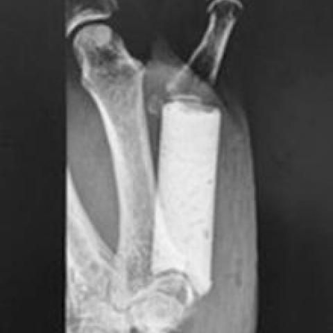
Plain radiographs demonstrated an expansile osteolytic lesion in the proximal epiphysis and diaphysis of the left 1st metacarpal bone (MC1). The cortex appeared markedly thinned. There was no surrounding sclerosis nor periosteal reaction (Fig. 1). CT confirmed a large lytic lesion with soft tissue attenuation, cortical thinning and destruction (Fig. 2). MR imaging showed bone marrow replacement with distal epiphyseal sparing. The lesion was of low signal intensity (SI) on T1-weighted images (WI) and intermediate to high SI on T2-WI. Moderate enhancement and soft-tissue extension was noted on contrast-enhanced images (Fig. 3). Bone scintigraphy demonstrated increased radionuclide uptake but absence of multifocality (Fig. 4). Surgical biopsy and subsequent histopathological examination revealed Giant Cell Tumour (GCT). Preoperative imaging (Fig. 5-6) 3 months later preceding MC1 resection (Fig. 7) and cement grafting (Fig. 8) showed marked increase of the lesion size. Histopathological findings of the resection specimen confirmed GCT without malignant degeneration.
Discussion
GCT of bone is a benign, but locally aggressive lesion with a tendency for local recurrence after resection. Metastasis is rare. Histologically, it is composed of multinucleated giant cells within a stroma of mononuclear cells. GCT account for approximately 5% of all primary bone tumours [1, 2]. It predominantly occurs between 20 and 50 years of age with a female predominance [1, 3, 4].
GCT of the bone commonly occur in the epi-metaphyseal region of long bones. The distal femur, proximal tibia and distal radius are commonly involved, with the spine and sacrum being less involved [3, 5]. The bones of the hand and foot are rarely involved, with a reported frequency of about 2% of all GCT. Metacarpal involvement is extremely rare [5].
Clinical presentation is usually nonspecific including pain, swelling, limited range of motion and pathological fractures [1, 5].
On conventional radiographs and CT, GCT is typically seen as an eccentric, epi-metaphyseal osteolytic lesion, with well-defined non-sclerotic border and extension underneath the subchondral articular bone [1-3, 5-7]. GCT may also show aggressive features consisting of poorly demarcated margins, cortical thinning and destruction and soft tissue extension [5]. CT may be useful in evaluating cortical bone integrity, absence of matrix mineralisation and demonstration of pathologic fracture [2, 4]. GCT of the hand tends to be less eccentric and more centrally located [7].
MR imaging can help determine the precise intramedullary and soft-tissue extent of the lesion [7]. Generally, GCT has a low-to-intermediate signal on T1-WI and a heterogeneous-high signal on T2-WI [2-4, 7]. However, due to intra-tumoural haemosiderin or fibrosis, the signal may be low on T2-WI [2]. The lesion enhances after intravenous gadolinium contrast administration, reflecting the increased vascular supply [6].
Bone scintigraphy may detect multifocality [4].
Extensive curettage or resection is the treatment of choice of GCT of the hand bones [2, 6, 7]. The combination of intraoperative cryogenic agents or methyl-methacrylate packing and resection has resulted in a recurrence rate of less than 10% [6]. For maintenance of the CMC function, resection is followed by bone reconstruction using an autogenous bone graft or allograft [2].
Although histology is mandatory for a definitive diagnosis, analysis of imaging characteristics of a lesion can be helpful in suggesting the correct diagnosis of a GCT. Epiphyseal extension and the low SI on T2-WI on MRI are useful signs in imaging characterisation of aggressive GCT even at rare localisations such as the metacarpal.
Written informed patient consent for publication has been obtained.
Differential Diagnosis List
Final Diagnosis
Giant cell tumour of the bone
Liscense
This work is licensed under a Creative Commons Attribution-NonCommercial-ShareAlike 4.0 International License.
Figures
Plain radiograph

CT findings


MR imaging findings


Bone scintigraphy

Plain radiograph (3 months later)

MRI findings (3 moths later)


Intra-operative

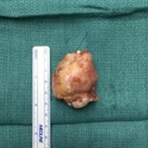
Post-operative
1. Imaging Findings
Based on the provided X-ray, CT, and MRI images, there is a notable osteolytic lesion in the proximal region of the first metacarpal bone of the left hand (first carpometacarpal joint). The lesion borders are mostly well-defined, but partial cortical thinning and mild expansion can be observed. The CT images show no obvious calcification or ossification within the lesion, which appears relatively homogeneous or septated with low density. On MRI, the area typically shows intermediate or slightly low signal intensity on T1-weighted images, and high or mixed signal intensity on T2-weighted images. Focal areas of low signal may suggest intratumoral hemorrhage or hemosiderin deposition. There is no evidence of extensive soft tissue invasion around the lesion, but adjacent soft tissue is slightly compressed.
2. Potential Diagnoses
- Giant Cell Tumor (GCT): Commonly occurs in individuals aged 20-50, with a slight female predominance. Often located at the epiphyseal/metaphyseal regions of long bones and crossing the subchondral bone. It presents as an osteolytic lesion with well-defined but aggressive borders. On MRI, it often shows mixed or high signal intensity.
- Osteoid Osteoma or Osteoblastic Lesions: Typically show prominent sclerosis or a sclerotic rim, often with a “reactive bone formation” on CT. This does not match the purely osteolytic nature and lack of definite sclerosis in this case.
- Aneurysmal Bone Cyst (ABC): More common in adolescents (ages 10-20), frequently affecting long bones and vertebrae. Often presents as an expansile osteolytic lesion with a “blown-out” appearance, sometimes septated. However, compared with giant cell tumors, certain features (like crossing the subchondral bone) are not as typical.
- Chondrosarcoma or Other Malignant Tumors: Often show more extensive soft tissue involvement or visible chondroid matrix (ring-like or arc-like calcifications). In this case, there is no distinct cartilage matrix calcification on imaging.
Considering the patient is a middle-aged woman with chronic localized pain and swelling, along with a typical osteolytic lesion extending to the articular surface on imaging, the top differential diagnosis is a giant cell tumor.
3. Final Diagnosis
Taking into account the patient’s age, symptoms, imaging findings, and the results of surgical pathology and histological examination, the most likely final diagnosis is:
Giant Cell Tumor of Bone
Although giant cell tumors are generally benign, they are locally aggressive and prone to recurrence. Therefore, both radiological and pathological assessments must be thoroughly evaluated. If uncertainty remains, a biopsy or postoperative pathological analysis can be performed for confirmation.
4. Treatment Plan and Rehabilitation
Treatment Strategy:
- Surgical Treatment: Wide curettage or resection is the preferred choice. If necessary, bone grafting (autograft or allograft) may be used to preserve joint function and prevent recurrence.
- Local Adjuvant Therapy: Intraoperative cryotherapy or filling the defect with bone cement (polymethyl methacrylate) can be employed to reduce the local recurrence rate.
- Other Treatments: If the lesion is extensive or surgical conditions are unfavorable, medication (such as osteoclast inhibitors) can be considered. However, surgery remains the mainstay in most cases.
Rehabilitation and Exercise Prescription (FITT-VP Principle):
- Initial Stage (Early Postoperative)
- Frequency: 1–2 times per day, with short practice sessions.
- Intensity: Gentle range-of-motion exercises within tolerable pain limits; avoid forceful gripping or twisting motions.
- Time: About 5–10 minutes each session, focusing on gentle movements of the fingers and wrist.
- Type: Assisted or active flexion/extension exercises of the finger joints and pinching movements. Use protective supports as needed to reduce stress.
- Progression: Gradual transition from passive to active exercises, combined with mild resistance training.
- Intermediate Rehabilitation
- Gradually increase range of motion and resistance training within a safe range, for example using a soft grip ball or elastic bands.
- Enhance wrist and thumb abduction/circumduction exercises to improve strength and flexibility.
- Late Rehabilitation
- Strengthen thumb opposition and finger coordination exercises, incorporating daily activities. Gradually add load (e.g., holding a pen, assisting in carrying objects).
- Resume normal work and sports activities progressively, ensuring local stability and healing.
- Precautions: If bone weakness or insufficient hand muscle strength is present, approach loading and resistance exercises with caution. In case of significant pain or swelling, re-evaluation and adjustment of the rehabilitation program are advised.
Disclaimer: This report is for medical information reference only and does not replace face-to-face diagnosis or a professional physician’s opinion. If there are any questions or the onset of new symptoms, please seek medical attention promptly.
Human Doctor Final Diagnosis
Giant cell tumour of the bone