Multiple sites of heterotrophic ossification in an infant mimicking multifocal osteomyelitis - Importance of SPECT/CT in paediatric imaging


Clinical History
This is a case of a two-month-old baby, with a previously drained left forearm abscess who was referred for a 2-phase bone scan in our facility for suspected multifocal osteomyelitis due to persistent fever despite negative cultures.
Imaging Findings
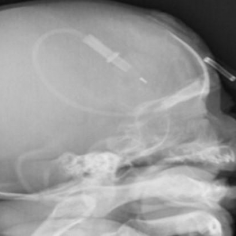
A 2-phase whole body bone scan was done and showed multiple sites of increased vascularity and osteoblastic activity respectively (Fig. 1). However, due to the limitations of planar imaging a single photon emission computed tomography (SPECT/CT) was ordered to accurately localise the abnormal foci of uptake. SPECT/CT localised the abnormal foci of uptake to soft tissue adjacent to the bones (Fig. 2 and 3). The CT only images showed these to be possible soft tissue calcification, which was confirmed on subsequent X-ray imaging (Fig. 4). The sites of abnormal soft tissue calcification corresponded to sites of previous trauma due to intravenous line insertions, incision and drainage (Fig. 5).
Discussion
Background
Heterotropic ossification (HO) is defined as the presence of bone in soft tissue, where it does not normally exist. [1] Most cases are acquired, usually following trauma, but could be congenital on rare occasions. [2] It is believed to result from transformation of primitive mesenchymal derived cells present in soft tissue into bone forming cells. [3]
Clinical perspective
The clinical diagnosis especially in the early stages of the disease could be difficult because of the non-specific symptoms, which includes pain, warmth, swelling and fever. [4] It is therefore difficult to distinguish it clinically from conditions like osteomyelitis that share similar symptoms. [5-8] This is where anatomical and functional imaging becomes important, as they can differentiate between the two conditions like in our case. The more commonly acquired form of HO may occur in virtually any form of musculoskeletal trauma. [1] In our case, the child experienced multiple sites of musculoskeletal trauma from previous drip insertions and the incision and drainage of the left forearm (Fig. 5).
Imaging perspective
A multiphase bone scan is used to aid in the diagnosis of HO and other conditions like osteomyelitis. [1] However, the limitations of planar imaging which include poor anatomic and spatial data has given rise to the use of SPECT/CT which has a more useful anatomic and spatial data, thereby improving localisation. [9] In our case, the SPECT/CT clearly showed that the abnormal foci was extraosseous and not in keeping with osteomyelitis. The soft tissue calcifications seen and confirmed on X-ray were in keeping with multiple sites of HO. Even though this patient is just 2 months old, literature has shown that a bone scan can be positive 2.5 weeks after injury and precede a positive radiography by 1-4 weeks. [10]
Outcome
Our findings were able to prevent further use of intravenous antibiotics in this case, knowing that 80% of patients with HO run a benign course. [4] The patient was discharged form the hospital about a week after imaging as fever had subsided, with a plan put in place for further management of HO if needed.
Take home message
Paediatricians should be aware that HO could occur as a complication of multiple drip insertions, considered as a possible diagnosis when osteomyelitis is being suspected. Appropriate imaging can differentiate the two and prevent wastage of resources on intravenous antibiotics and prolonged hospital stay. This is the first case of paediatric HO in our department.
Written informed patient consent for publication has been obtained.
Differential Diagnosis List
Final Diagnosis
Multiple sites of heterotrophic ossification
Liscense
This work is licensed under a Creative Commons Attribution-NonCommercial-ShareAlike 4.0 International License.
Figures
X-ray


Photos


2-phase bone scan


SPECT/CT


SPECT/CT


Medical Imaging Analysis Report
1. Radiological Findings
The patient is a 2-month-old male infant who previously underwent incision and drainage for a left forearm abscess. He continued to have a fever, though all bacterial cultures remained negative. Multiple-phase bone scans and X-ray examinations revealed the following:
- Focal calcifications on the left forearm and in multiple soft tissue areas, appearing as high-density shadows or bone-like structures within soft tissue.
- No significant signs of bone destruction; the cortical integrity of the bones is intact.
- CT/SPECT further indicates that these calcifications are located within soft tissue compartments, rather than within bone, suggesting abnormal ossification in soft tissue.
- Multiple-phase bone scans show “hot spots” localized in soft tissue regions with a clear boundary from bony structures. Radiotracer accumulation is consistent with heterotopic ossification (HO) on imaging.
- Head X-rays and scans do not show obvious erosion of the skull or any fracture displacement. Close attention should be given to potential soft tissue injuries around the head, such as areas affected by cannulation/fixation devices.
2. Possible Diagnoses
Based on the above radiological findings and the infant’s clinical history, the following diagnoses should be considered:
- Multiple Heterotopic Ossification (HO)
- The infant has undergone IV infusions and incision and drainage in the forearm, leading to multiple soft tissue injuries or interventions.
- Bone-like lesions appearing in soft tissue with clear margins, and no destruction of the actual bone, align with the mechanism of HO.
- Bone or Soft Tissue Infection (e.g., Osteomyelitis, Soft Tissue Abscess)
- Persistent fever previously raised this possibility, but culture results were repeatedly negative, and imaging does not show typical bone involvement or cortical destruction.
- In cases of bone infection, bone scans typically indicate radiotracer uptake in the bone along with bone destruction or periosteal reaction, which are not present here.
- Rare Congenital Ossification Disorders or Pediatric Tumors
- Given the patient’s age (2 months), congenital bone formation disorders or tumoral calcifications (e.g., osteochondroma, osteosarcoma) must be excluded. However, clinical presentation and imaging findings are not typical for these conditions.
3. Final Diagnosis
Taking into account the infant’s age, history of trauma or invasive procedures (IV infusions, surgical drainage of the forearm), persistent fever with negative cultures, and multi-modality imaging findings (bone scan, X-ray, SPECT/CT), the most likely diagnosis is:
Multiple Heterotopic Ossification (HO).
HO generally has a favorable prognosis, and imaging has ruled out osteomyelitis or other aggressive pathologies. If suspicious symptoms or signs worsen clinically, additional follow-up imaging (MRI or repeat bone scans) can be conducted for ongoing evaluation.
4. Treatment and Rehabilitation Plan
- Treatment Strategy
- Typically, there is no need for aggressive antibiotic therapy: Since bone infection has been excluded, the empirical or extended use of IV antibiotics is unwarranted.
- Control inflammation and pain: In cases of significant local inflammation, pediatric-appropriate doses of NSAIDs may be considered for a short duration. If there is minimal pain, no medication may be needed.
- Regular follow-up: Most HO lesions either resolve spontaneously or stabilize. Periodic imaging evaluations are necessary to monitor for any progressive enlargement or joint involvement.
- Surgical intervention may be considered if HO growth is substantial, compresses neurovascular structures, or severely affects function. However, in infants, it is prudent to minimize surgical trauma.
- Rehabilitation/Exercise Prescription
- Frequency: Several short sessions per day involving gentle stretching or passive joint movements. Movements should be performed gently and in moderation.
- Intensity: Movements should not cause significant pain or distress. Care must be taken with the affected limb to avoid excessive traction and repeated irritation at the wound or ossification sites.
- Duration: Each session can last approximately 5-10 minutes, adjusted according to the infant’s tolerance.
- Methods: Under the guidance of a qualified caregiver or professional, gentle manipulation of all four limbs, including light passive movements of the elbow and wrist, is encouraged. Natural active movements should be promoted within comfortable limits.
- Progression: If soft tissue swelling subsides and the infant shows no notable discomfort when calm, the range of motion and duration of exercises can be gradually increased. Observe local and systemic reactions closely.
Since the patient is only 2 months old, any exercise prescription should be implemented by caregivers or rehabilitation specialists, focusing on gentle active/passive joint mobility and basic infant massage:
Rehabilitation strategies for infants require comprehensive assessment of cardiopulmonary function and skeletal development. For this case, the focus is on preserving local soft tissue and joint function while avoiding further injury or aggravating abnormal ossification.
Disclaimer: This report is based on the limited medical records and imaging provided. It is for reference only and cannot replace in-person consultation or the professional judgment of a licensed physician. If there are any concerns or changes in condition, please seek prompt medical attention and discuss with a qualified healthcare provider.
Human Doctor Final Diagnosis
Multiple sites of heterotrophic ossification