


Patient with left dorsolateral forefoot pain. Six months of evolution. Without trauma or functional limitations.
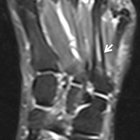
The radiographic examination showed a narrowed intermetatarsal space with osseous hypertrophy and bridging between the bases of the 3rd and and 4th metatarsals (Fig. 1). CT also noted areas of bone degenerative appearance with sclerosis and osseous proliferation (Fig. 2, 3). MRI showed this anomalous joint with intermediate signal intensity in T1 and T2-weighted images with osseous oedema (Fig. 4).
Coalition occurs when two or more bones are joined by a bar or bridge between them.
Histologically it may be fibrous (syndesmosis), cartilaginous (synchondrosis), or osseous (synostosis). The incidence of tarsal coalition is classically thought to be approximately 1%; however, many feel that this is an underestimate as only the symptomatic cases come to attention.
Most cases are present in the tarsus, mainly in the calcaneonavicular and talocalcaneal joints causing chronic pain in the hindfoot [1]. However, intermetatarsal coalitions in the forefoot are extremely rare, with only a small number of cases reported. Most occur between the base of the fourth and fifth metatarsals and, less commonly, between the first and second metatarsals [4]. Coalitions can be congenital (originate in a failure of embryonic mesenchymal differentiation presenting autosomal dominant hereditary pattern, mainly in bilateral cases) or acquired (osteoarthritis, rheumatoid arthritis, infection, trauma, diabetes, neoplasia or iatrogenic origin) [2, 3].
Pain is the most common presenting symptom, followed by bone deformity and stiffness of the joints [5].
The radiographic findings are more evident in the osseous type, where a bridge and cortical continuation between the involved metatarsal bones is observed. In non-osseous types, the two involved metatarsal bones can demonstrate abnormal narrowing and irregularity of the involved joint space (Fig. 1), which may radiographically appear to be degenerative changes. This is important in young patients where you would not expect to see any degenerative disease [6].
CT findings vary according to the type of coalition. Osseous coalitions presents anomalous bone continuity of two or more bones, while in non-bone coalitions, as in our case, they usually present an abnormal narrowing of the joint space or pseudoarticular appearance (Fig. 2 and 3).
MRI also shows bone marrow across the fused articulation in the osseous type.
Non-osseous coalitions (Fig. 4, 5) demonstrate on MRI narrowing of the joint space, and irregularity of the bone interface. Soft tissue and bone marrow oedema can be seen commonly in symptomatic patients. Cartilaginous coalitions will demonstrate signal intensity similar to fluid or cartilage, and fibrous type will demonstrate low-signal intensity on all sequences. When this differentiation is not so characteristic, the coalition is categorised as non-osseous.
The therapy includes surgery and conservative treatment [6, 7].
In conclusion, it is important to be able to recognise this entity because, when it is symptomatic, it can be a cause of metatarsalgia-like pain, in young and adults patients.
Written informed patient consent for publication has been obtained.
Proximal non-osseous coalition between the third and fourth metatarsals
This work is licensed under a Creative Commons Attribution-NonCommercial-ShareAlike 4.0 International License.


X-ray, CT, and MRI images reveal that the 4th and 5th metatarsal bases of the patient’s left forefoot are abnormally close, with marked joint space narrowing, irregular cortical margins, or a tendency toward partial fusion. The 3D CT reconstruction indicates a sign of non-osseous bridging (suspected fibrous or cartilaginous tissue) between the two metatarsals. MRI further demonstrates joint space narrowing in this area, with mild edema signals in the local bone and surrounding soft tissues, suggesting changes likely caused by relatively chronic inflammation or frictional irritation.
Taking into account the absence of trauma, the lesion being localized to the bases of the 4th and 5th metatarsals, and imaging findings suggesting bridging, the presentation is most consistent with an intermetatarsal coalition.
Considering the patient’s age, symptoms (lateral dorsal forefoot pain in the left foot), and imaging findings of a non-osseous bridge between the 4th and 5th metatarsal bases, the most likely diagnosis is: Non-osseous Intermetatarsal Coalition of the 4th and 5th Metatarsals in the Left Foot (Intermetatarsal Coalition).
If necessary, further confirmation can be achieved by correlating the severity of clinical symptoms with other tests, such as diagnostic local anesthetic block or intraoperative exploration.
Rehabilitation training should follow a gradual approach (FITT-VP principle) and be individualized:
Since the patient already exhibits structural changes in the forefoot, it is crucial to monitor the load on the metatarsal joints and their stability. Should localized pain, swelling, or discomfort intensify during training or daily activities, consult a physician or contact a rehabilitation therapist promptly to adjust the program.
Disclaimer: This report is a reference analysis based on the currently provided information and does not replace in-person consultation or professional medical advice. If you have any questions or if symptoms worsen, please seek assistance from a specialist promptly.
Proximal non-osseous coalition between the third and fourth metatarsals