Kienböck’s disease:Role of MRI in interpretation and management to prevent progression of the disease


Clinical History
A 34 years old male driller by profession presented to our radiology department with pain in
right wrist .Pain had worsened over a period of six months and patient was having difficulty in doing daily works
Imaging Findings
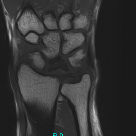
X ray of right hand revealed significant sclerosis of lunate bone with disuse osteopenia in adjacent carpal bones. MR images through the right wrist showed collapse of the lunate with diffuse abnormal T1 hypointense signals seen in it. Abnormal signals were also seen involving the capitate and scaphoid bone (proximal to the waist of scaphoid). Few abnormal signals with focal areas of cartilaginous thinning and tiny cysts were identified involving the articular and sub articular surfaces of all the carpal bones, distal radius and the ulna. Overall findings were consistent with osteonecrosis of the lunate bone.
Discussion
Kienböck’s disease also referred to as osteonecrosis of lunate bone or called lunatomalacia or avascular necrosis of the lunate bone, is a rare condition in which lunate bone, loses its blood supply leading to avascular necrosis. [1] Its causes are multifactorial, commonly affecting men between the ages of 20 and 40 years. Negative ulnar variance results in increased shear stress to the lunate bone which might be a risk factor to develop lunate necrosis through still unknown mechanisms. Regarding lunate pressure it has been debated that increased intraosseous pressure caused by venous stasis (i.e. in extension) might be another risk factor or cause for lunate necrosis. [2].
CLINICAL PERSPECTIVE: In early stages of the disease patient may present with pain, edema and limited wrist motion. With disease progression bone destruction and collapse occurs resulting in secondary wrist osteoarthritis. Early diagnosis and treatment help in prevention and progression of necrotic changes and bone collapse.
IMAGING PERSPECTIVE: The management of Kienböck’s disease is highly dependent on the stage of the disease based on the Lichtman classification system. MRI (Magnetic resonance imaging) is helpful early in the disease when plain radiographs are not helpful however bone contusion or acute fracture must be ruled out by adequate clinical history for the differential of a Kienböck's disease stage I. Contrast-enhanced MRI is important for assessment of the degree of necrotic tissue and for the treatment of stage II and III A disease. MRI with contrast is often not necessary in stages I, III B, III C, or IV because in such cases degree of necrosis does not usually change treatment. [3]
OUTCOME: There is lack of consensus among hand surgeons about treatment of disease. For patients with ulnar-minus variance and stage I disease immobilization is recommended. For stages II or III A disease with ulnar-minus variance, an equalization procedure is attempted. In stage III B disease, Scapho-trapezial-trapezoidal (STT) arthrodesis is done to restore carpal stability and prevent further degeneration. In stage IV disease, proximal row carpectomy (PRC) or wrist arthrodesis is done. [4] Take home message is that radiology plays an important role in the diagnosis and classification of the disease process. MRI plays a vital role in the early diagnosis of stage I of the disease process and is superior in sensitivity and specificity to bone scan in early detection and prevention of disease progression.
Written informed patient consent has been taken for publication
Differential Diagnosis List
Final Diagnosis
Kienböck’s disease Litchman stage IV
Liscense
This work is licensed under a Creative Commons Attribution-NonCommercial-ShareAlike 4.0 International License.
Figures
CORONAL MRI RIGHT HAND

X RAY RIGHT HAND AP VIEW

MRI RIGHT HAND CORONAL T1W IMAGES
X RAY RIGHT HAND LATERAL VIEW

MRI RIGHT HAND CORONAL STIR IMAGES

Medical Analysis Report
1. Radiological Findings
Based on the provided wrist X-ray (PA and lateral views) and MRI images, the main findings are as follows:
• The PA view of the wrist X-ray shows slightly abnormal density in the lunate region, with sclerosis and morphological changes compared to the surrounding bones. In some cases, there may be signs of lunate collapse.
• On MRI sequences (especially T1 and T2-weighted images), there is an area of abnormal signal within the lunate, presenting as low or mixed signal intensity, indicating local vascular compromise or necrosis.
• There is no clear acute fracture line, but the lunate shows characteristic structural and signal changes consistent with vascular compromise and necrotic processes.
Additionally, in some images, there is a mild ulnar-minus variance that may be contributing to increased compressive stress on the lunate. No extensive bony destruction of the surrounding joint surfaces is noted yet, but persistent disease progression may lead to secondary osteoarthritis changes.
2. Possible Diagnoses
-
Kienböck’s Disease (lunate avascular necrosis):
The most characteristic feature is aseptic necrosis of the lunate, commonly seen in patients aged 20–40 years, often associated with ulnar-minus variance or chronic wrist stress. Imaging typically shows lunate signal abnormalities and sclerosis, and in later stages, lunate collapse.
-
Bone Contusion or Occult Fracture:
If there is a history of trauma or repetitive vibrating stress, a bone contusion may present with similar MRI signal abnormalities. However, these usually improve over several weeks or months, and there is often a clear history of trauma or stress-related injury.
-
Other Vascular Supply Issues of the Carpal Bones (rare):
Similar vascular supply issues may occasionally affect other carpal bones such as the scaphoid, but the clinical and imaging findings differ. In this case, the typical pattern is more consistent with lunate necrosis.
3. Final Diagnosis
Considering the patient’s age, occupational factors (commonly linked with sustained vibration or pressure load), wrist pain symptoms, and imaging findings,
the most likely diagnosis is Kienböck’s Disease (aseptic necrosis of the lunate).
If further confirmation of the necrosis range or assessment of surgical indications is needed, contrast-enhanced MRI or other imaging studies may be performed to evaluate the degree of vascular compromise within the lunate.
4. Treatment Options and Rehabilitation Plan
(1) Treatment Strategy
• Conservative Treatment: Suitable for early-stage disease (Stage I) or patients with limited involvement. Typical recommendations include immobilization of the wrist or casting/splinting, reducing weight-bearing and excessive wrist movements.
• Surgical Treatment: Depending on the Lichtman staging and the specific situation:
– For Stage II or IIIA with ulnar-minus variance, ulnar shortening or radial lengthening osteotomies can be considered to reduce compressive stress on the lunate;
– For Stage IIIB or later, if the disease has led to significant lunate collapse, a Scapho-Trapezoid-Trapezium (STT) joint fusion or other reconstructive procedure may be performed;
– For Stage IV with obvious osteoarthritic changes, a proximal row carpectomy or total wrist fusion can be considered.
(2) Rehabilitation and Exercise Prescription
After the acute immobilization or surgical intervention, a gradual rehabilitation program for wrist function and muscle strength is needed, following the FITT-VP principle:
• Frequency: Initially, perform simple wrist exercises (fist making, flexion/extension) 1–2 times a day; in later stages, increase to 2–3 times a day, including resistance training.
• Intensity: Begin with low-intensity, no-resistance exercises such as active wrist movements and gentle squeezing of a grip ball. As pain and discomfort subside, gradually introduce light resistance training.
• Time: Start with 5–10 minutes per session and may extend to 15–20 minutes based on tolerance and symptom response. Avoid overstressing the wrist if it causes pain escalation.
• Type: Include wrist flexion/extension, rotation, and light resistance exercises. These can be supplemented with exercises for the fingers and forearm muscles, such as using a grip strengthener or light dumbbells.
• Progression: Depending on wrist mobility and pain levels, increase intensity or duration every 1–2 weeks. If significant swelling or pain occurs, reduce the load and consult your physician.
• Special Considerations: If the patient has fragile bones or is in the postoperative recovery phase, training should be conducted under the guidance of a professional rehabilitation therapist to ensure wrist stability and safety.
5. Disclaimer
This report provides a preliminary analysis based solely on the given clinical and imaging information, and cannot replace an in-person consultation or professional medical advice. If you have further symptoms or concerns, please contact a healthcare professional or visit a hospital for more comprehensive and individualized diagnosis and treatment recommendations.
Human Doctor Final Diagnosis
Kienböck’s disease Litchman stage IV