Panner's disease


Clinical History
A 7-year old youth soccer player presented with periodic pain and slight swelling of his right elbow since two months, without previous trauma. There was a limited range of motion in the elbow.
Imaging Findings
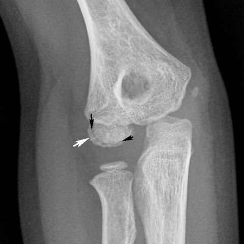
Conventional radiography showed joint effusion, irregular contours of the capitellum, increased bone density and a thin radiolucent line in the subchondral bone (Figures 1a and 1b). Follow-up with Cone Beam CT 2 months later depicted a crescent with vacuum phenomenon in the subchondral bone (Figures 2a and 2b), corresponding to the radiolucent line seen on radiographs.
Subsequent MRI revealed intact articular cartilage of the capitellum, predominantly low signal of the capitellum on both T1- and PD weighted images in keeping with sclerosis, and bone marrow edema in the adjacent humeral bone on FS PD weighted images (Figures 3a, 3b, 3c and 3d).
Discussion
Panner’s disease represents an osteochondrosis of the humeral capitellum. Osteochondroses affect epiphyses or epiphyseal equivalents and their progression ceases with skeletal maturity of the affected part [1]. Panner’s disease occurs most frequently in boys under the age of 11 [2, 3].
Microtraumata from repetitive valgus stress and increased axial load are incriminated as the main pathogenetic factors, typically occurring in throwing sports and gymnastics respectively [2].
This may lead to disruption of vessels that supply the nucleus of the capitellar ossification centre, leading to ischemia, resulting in disturbed endochondral ossification [2].
Clinical presentation consists of pain, stiffness, swelling and a limited range of motion – especially an extension deficit, flexion deficit is less common [2]. The pain is activity-related [2].
Conventional radiographs may show an irregular contour, sclerosis, subchondral radiolucency, fragmentation and flattening of the capitellum [2, 3].
As the radiographical changes may be subtle, a low dose (Cone Beam) CT may be beneficial because of its greater sensitivity for detection of subtle changes and characterization of a subchondral vacuum phenomenon [4].
The radiolucent subchondral crescent represents gas accumulation in a subchondral fissure, which indicates osteonecrosis and impending articular collapse [4, 5]. This vacuum-phenomenon disappears when repair and re-ossification occurs [4].
MRI shows heterogeneous signal intensity due to a variable degree of sclerosis, vacuum phenomenon and bone marrow edema in the ossified part of the capitellum, while the overlying cartilage is normal [2, 4]. The vacuum phenomenon has a similar low signal as the sclerotic changes and is therefore difficult to detect on MRI.
The majority of patients can be successfully treated conservatively, without serious morbidity [2].
The differential diagnosis of Panner’s disease consists of osteochondrosis dissecans (OCD), which is seen in the second decade of life, when the ossification of the capitellum is complete [3, 5]. OCD affects both bone and overlying cartilage and may be complicated by intra-articular loose body formation and has an overall poorer outcome [2, 3]. MRI may detect cartilage defects, therefore it will be useful for distinguishing between Panner’s disease and OCD.
Our patient was treated conservatively, with rest, avoidance of weight bearing and of leaning upon the right arm, promotion of passive movement and antiphlogistic medication. After six months there was a significant clinical improvement and a partial regression of radiological abnormalities.
Written informed patient consent for publication has been obtained.
Differential Diagnosis List
Final Diagnosis
Panner’s disease.
Liscense
This work is licensed under a Creative Commons Attribution-NonCommercial-ShareAlike 4.0 International License.
Figures
Conventional radiograph of the right elbow, lateral and anteroposterior view

Cone Beam CT of the right elbow


MRI of the right elbow,T1 and FS PD


Medical Analysis Report
1. Imaging Findings
Based on the provided X-ray, CT, and MRI images, the following findings are observed:
- X-ray Findings: The humeral capitellum (i.e., the distal lateral aspect of the humerus) appears irregular in shape, with mild collapse or flattening of the local margin. Areas of sclerosis (increased density) and a subchondral radiolucent line (vacuum sign) can be observed.
- CT Findings: A gas-density shadow (vacuum sign) within the subchondral fissure of the humeral capitellum is noted, with mixed high-density sclerosis and low-density fragmentation. The outline is slightly irregular.
- MRI Findings: On T1- and T2-weighted images, the humeral capitellum shows heterogeneous signal, suggesting bone sclerosis and partial bone marrow edema. The cartilage layer remains essentially intact without obvious cartilage defects or loose bodies. The vacuum sign appears as low signal on MRI, making it difficult to distinguish from sclerosis zones, but mild edema can be seen in the adjacent areas.
2. Possible Diagnoses
Considering the patient’s age (7 years), symptoms (chronic dull pain, restricted range of motion), and imaging features (sclerosis, fragmentation, vacuum sign of the humeral capitellum, and intact cartilage), the primary possible diagnoses include:
- Osteochondrosis of the Humeral Capitellum (Panner’s Disease): Commonly seen in children under 11 years old, especially those engaged in repetitive throwing or weight-bearing sports (such as gymnastics or pitching) leading to microtraumatic stress. Imaging typically shows capitellar irregularity, sclerosis, and fragmentation. The cartilage remains relatively unaffected due to the ongoing plasticity, so the articular surface typically does not show major defects.
- Osteochondritis Dissecans (OCD): More common in slightly older age groups (usually over 12 years), involving both cartilage and subchondral bone. There may be subchondral fractures, partially detached or free fragments, and possible intra-articular loose bodies. Imaging findings often indicate discontinuity or separation of the cartilage surface.
- Other Joint Pathologies: Though less common, conditions such as infection (osteomyelitis) or post-traumatic avascular necrosis should be considered. Distinguishing factors include inflammatory markers and specific imaging features.
3. Final Diagnosis
Taking into account the patient’s age, clinical symptoms (chronic elbow discomfort, restricted motion, typical history of throwing or weight-bearing activities), and imaging findings (subchondral sclerosis, fragmentation, vacuum sign in the humeral capitellum, with no major cartilage damage), the most likely final diagnosis in this case is:
Osteochondrosis of the Humeral Capitellum (Panner’s Disease).
4. Treatment Plan and Rehabilitation
For Panner’s disease, since the patient’s skeletal system is still developing, most cases can be managed with conservative treatment and a rehabilitation plan as follows:
- Rest and Immobilization: Advise the patient to avoid activities that increase stress on the humeral capitellum, such as throwing, gymnastics, prolonged weight-bearing, or supporting body weight on the arms. Short-term use of an elbow brace or rest can help alleviate pain and expedite healing.
- Medication: During acute phases or when pain is significant, a short course of non-steroidal anti-inflammatory drugs (NSAIDs) can be used to reduce pain and inflammation.
-
Rehabilitation Exercises: After acute symptoms subside, a progressive therapy program under the guidance of a qualified rehabilitation or sports therapist is recommended:
-
Phase I (Symptom Relief Phase):
• Frequency: 1-2 times per week
• Intensity: Low-intensity passive/active joint mobility exercises, such as gentle flexion-extension of the elbow and forearm rotation
• Duration: 10-15 minutes per session
• Method: Performed without provoking significant pain; may be combined with heat therapy or physiotherapy to relieve muscle tension. -
Phase II (Functional Recovery Phase):
• Frequency: 2-3 times per week
• Intensity: Gradually introduce resistance exercises, mild grip training, and wrist-based exercises, avoiding excessive impact on the elbow joint
• Duration: 15-20 minutes per session
• Method: Light resistance band exercises focusing on the triceps and forearm muscle groups (isometric or low-load isotonic). -
Phase III (Strengthening Phase):
• Frequency: Approximately 3 times per week
• Intensity: Gradual return to light throwing or striking motions, following a cautious, stepwise progression
• Duration: 20-30 minutes per session
• Method: Include core stability and shoulder strengthening exercises to reduce elbow stress.
-
Phase I (Symptom Relief Phase):
- Exercise Progression (FITT-VP Principle): Adjust the frequency, intensity, time, type, volume, and progression of exercises according to the patient’s subjective pain level and objective assessments of joint range of motion and muscle strength.
- Surgical Intervention: Considered only if conservative treatment fails or if significant cartilage damage or loose bodies are present. However, the prognosis for Panner’s disease is typically good, and most cases do not require surgery.
Disclaimer: This report is a reference-based analysis of the information provided and does not replace an in-person consultation or professional medical diagnosis and prescription. If you have any concerns or worsening symptoms, please seek medical attention promptly.
Human Doctor Final Diagnosis
Panner’s disease.