Kissing spine syndrome: An often underdiagnosed cause of back pain


Clinical History
A 53-year-old-male patient with history of long standing low-back pain for which he had previous lumbar spine X-ray. Patient reports recent increase in back pain with some gait disturbance. On examination tender L3 to S1 vertebral level and positive SLR (straight leg test). MRI lumbosacral spine was suggested to rule out any nerve root compression.
Imaging Findings
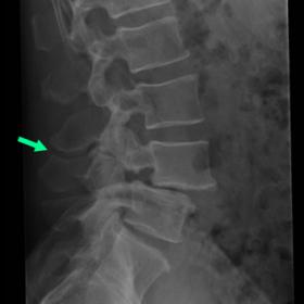
X-ray lumbar spine lateral view (Fig. 1) done few months before. The MRI showed grade 1 anterolisthesis of L4 on L5 vertebra, with underlying pars interarticularis defect and narrowing of interspinous distance at L3-L4 level (green arrow). Sagittal T2 and STIR sequences (Figs. 2,3) show interspinous bursal fluid at L3-L4 level (blue arrows) which can be traced anteriorly to a posterior epidural cyst measuring 20 x 13mm (yellow arrows). In addition, subtle oedema and tiny cyst seen in L4 spinous process in sagittal STIR image. Axial T2 (Fig. 4) shows canal compromise and indentation of cauda equina nerve roots seen due to posterior epidural cyst (green arrow). Features were consistent with Baastrup’s disease. Other findings include facet joint arthropathy at L3-L4 level, grade 1 anterolisthesis of L4 on L5 vertebra with underlying pars interarticularis defect and end plate marrow oedema at L4-L5 level.
Discussion
Baastrup’s disease (also known as kissing spine syndrome) is a relatively common, yet underdiagnosed cause of back pain [1]. It was named after Christian Ingerslev Baastrup in 1933, who initially described it as a condition where the adjacent spinous process of vertebra are closely approximated in the setting of degenerative spine disease [2,3]. Typically, patients complain of midline lumbar back pain which is aggravated by extension and relieved by flexion or symptoms related to canal compromise [1,3,4,5]. The disease commonly affects the lumbar spine and mostly involves a single level [1,4]. The interspinous ligament is responsible for maintaining the sagittal stability of the spine. The pathology involves progressive weakening of the ligament as a result of stretching caused by shearing force applied to it with movement (loading and ambulation).The progressive weakness of the ligament with increase in bursal fluid due to friction and associated inflammatory change between the spinous processes leads to formation of fluid clefts and subsequently epidural cysts [6,7]. The cysts develop in the weakest area, which is the ventral area near the posterior epidural space. Weakening of the interspinous ligament leads to spinous process approximation, sagittal instability and anterolisthesis [8,9,10]. Although this disease has been shown previously as a separate entity, independent of other pathology [3,11], it can be associated with degenerative disc disease, spondylolisthesis and spondylosis as seen in our case report [4]. Imaging plays a vital role in identifying this entity and differentiating this from other causes of back pain. Radiograph and CT often show close approximation and reactive sclerosis of the hypertrophied spinous process [12]. MRI is best suited for an early diagnosis as it has been noted that interspinous bursitis may precede osseous changes of spinous process seen on X-rays [6]. MRI reveals a spectrum of findings including spinous process oedema, sclerosis and subchondral cyst, interspinous ligament fluid cleft, posterior epidural cyst, ligamentum flavum thickening and anterolisthesis of caudal vertebra in the levels involved [5]. Though PET-CT is not routinely used for diagnosing this condition, FDG (flurodeoxy-glucose)-uptake may be seen at interspinous ligament and spinous process level indicating the site of active inflammation [13]. Management includes conservative approach with physiotherapy and anti-inflammatory medications, percutaneous injection of steroids and surgical decompression for canal compromise [3,14-16]. Written informed patient consent for publication has been obtained.
Differential Diagnosis List
Final Diagnosis
Baastrup’s disease with posterior epidural cyst causing canal compromise
Liscense
This work is licensed under a Creative Commons Attribution-NonCommercial-ShareAlike 4.0 International License.
Figures


Medical Imaging Analysis Report
I. Imaging Findings
Based on the lateral lumbar spine X-ray provided by the patient, a narrowing of the distance between adjacent spinous processes can be observed, with local evidence of noticeable hyperostosis and sclerosis suggestive of the “kissing” phenomenon of the spinous processes. On MRI T2-weighted images, distinct fluid-like or cystic signals are visible in the interspinous space posterior to the lumbar spine, partially adjacent to the dural sac, potentially causing mild compression or reduction of the dural sac. Degenerative changes are seen in the intervertebral discs and adjacent joint surfaces, with some intervertebral spaces showing slight narrowing and local ligament hypertrophy or thickening. These intermittent cystic changes and the “touching” sign of the spinous processes are consistent with Baastrup’s syndrome (also known as “kissing spine” or “kissing spinous process syndrome”).
II. Potential Diagnoses
-
Baastrup’s Syndrome (Kissing Spine Syndrome)
Cause: Long-term lumbar spinal degeneration leading to abnormal proximity or friction between spinous processes, prompting bursa-like changes, inflammation, and pain in the interspinous space and posterior area. Radiological signs include spinous process enlargement, deformation, and cystic or fluid signals near the spinous processes. -
Degenerative Disc Disease
Cause: Disc degeneration can manifest as narrowed intervertebral spaces, marginal osteophytes on vertebral bodies, and varying degrees of signal changes, often accompanied by low back pain. However, mere degenerative changes rarely present obvious “touching” spinous process signs. -
Spondylolisthesis
Cause: Degenerative changes or structural instability result in anterior/posterior slippage of the vertebrae. Some patients show spinous process changes, but if there is no significant slip or instability, Baastrup’s syndrome should be considered separately. -
Epidural Cyst or Posterior Spinal Canal Cyst
Cause: Degenerative changes or local inflammation may lead to fluid protrusion behind the spine on MRI. Other intraspinal space-occupying lesions should be excluded.
III. Final Diagnosis
Considering the patient’s age, history of chronic low back pain, and imaging findings of pronounced interspinous alterations along with mild dural sac compression, the most likely diagnosis is:
Baastrup’s Syndrome (also known as Kissing Spine Syndrome)
This condition often coexists with degenerative changes in the lumbar spine, such as disc degeneration, osteophyte formation, and ligament hypertrophy, which aligns with this patient’s presentation. If clinical symptoms worsen or there is marked neural compression later, further evaluation with CT or assessment of spinal stability may be necessary to decide on treatment strategies.
IV. Treatment Strategy and Rehabilitation Plan
1. Conservative Treatment
- Pain relief and anti-inflammatory therapy: Use of NSAIDs and analgesics to alleviate local inflammation and pain.
- Physical therapy: Warm therapy, ultrasound therapy, and muscle massage to relieve local muscle spasms and improve blood circulation.
- Exercise training: Strengthening of core and lumbar back muscles to enhance spinal stability.
2. Interventional and Surgical Management
- Interventional injections: If pain is significant and conservative therapy yields limited results, consider interspinous or epidural injection of corticosteroids and local anesthetics to relieve pain and local inflammation.
- Surgical decompression: For cases of obvious spinal canal stenosis or nerve root compression, surgery may be considered, including partial resection of hypertrophic spinous processes or decompression and fusion procedures.
3. Example of a Rehabilitation/Exercise Prescription (Following the FITT-VP Principle)
- Frequency: 3–5 times per week, depending on the patient’s general condition.
- Intensity: Start with light to moderate intensity (e.g., mild resistance training, avoiding excessive extension) within a range that does not trigger significant pain or only causes mild discomfort.
- Time: 20–30 minutes per session, which can be divided as needed. For instance, begin with 5 minutes of core contraction exercises, rest, then proceed with 10 minutes of lumbar back muscle exercises.
- Type: Focus on core stability, lumbar back muscle strengthening, and balance training, supplemented by low-impact aerobic exercises (e.g., elliptical trainer or brisk walking on flat ground).
- Progression: Once the patient can complete the current stage pain-free, gradually increase exercise volume, such as extending session length to 30–40 minutes or modestly increasing resistance or repetitions.
- Volume & Individualization: Total training volume and methods should be adapted according to the patient’s tolerance, cardiopulmonary function, and bone health status (avoiding high-impact moves if there is a risk of osteoporosis).
Disclaimer
This report is based solely on the current imaging and medical history for reference purposes and does not replace an in-person consultation or professional medical advice. If there are any questions or changes in condition, please seek medical attention promptly for further personalized guidance.
Human Doctor Final Diagnosis
Baastrup’s disease with posterior epidural cyst causing canal compromise