Atypical sequence of elbow ossification with asymmetric appearance of proximal radius epiphyses: A Pathology mimicker


Clinical History
A 9-year-old boy presented with a painful left forearm and elbow after falling down yesterday. On examination there was diffuse swelling over the left forearm and elbow associated with tenderness especially at the mid-forearm. The range of motion in the forearm and elbow was guarded.
Imaging Findings
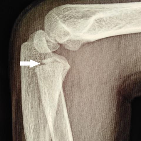
Anterior-posterior and lateral radiographs of the left forearm showed a uni-cortical fissure fracture of the mid-shaft of the radius (Fig. 1). Additionally, the anterior-posterior and lateral radiographs of left elbow showed that the proximal radius epiphysis was non-ossified. At the same time the medial epicondyle epiphysis was completely ossified in both the anterior-posterior and lateral views (Fig. 2). Contrastingly, radiographs of the normal right elbow showed a well-ossified proximal radius epiphysis. Likewise, the medial epicondyle epiphysis was completely ossified (Fig. 3).
Discussion
Radiographic evaluation of the acutely injured paediatric elbow presents a particularly challenging problem for emergency department physicians and radiologists alike. Misreading plain radiographs of the acutely traumatised elbow in children can lead to misdiagnosis and failure to institute treatment in a timely fashion with subsequent functional disability [1,2]. Contrastingly, failure to recognise normal anatomic variants simulating pathology may result in groundless over-investigations and over-treatment.
The radiologic anatomy of the growing child is complicated. The timing of appearance of elbow secondary ossification centres shows considerable variations regarding age, gender and race. Likewise, the symmetrical appearance of elbow ossification centres can show diversity. The chronological order of appearance of the ossification centres is an important guide to an accurate interpretation of paediatric elbow radiographs in the acute setting [3,4]. This chronological order of appearance of the elbow ossification centres has a general tendency to follow this sequence; capitellum, radial head, medial epicondyle, trochlea, olecranon, and lateral epicondyle collectively known as (CRITOL). They ossify at 1, 5, 10, and 11 years, respectively. Although this chronological order is a fairly reliable radiographic finding, it should be contextualised with race, gender and age [4-9]. In some studies the reported age of appearance of the medial epicondyle epiphysis was earlier than the proximal radius epiphysis. Similarly, the reported age of appearance of the olecranon was earlier than the trochlea epiphysis [4,5,6,8,9]. The incidence of normal variances was reported to be higher in girls approaching near statistical significance [9].
Generally, the radial head epiphysis ossifies before or simultaneously with the medial epicondyle epiphysis [1,3]. Our patient exhibited an atypical but normal sequence of ossification. The radiographs of the injured left elbow showed a well-ossified medial epicondyle epiphysis while the radial head epiphysis was non-ossified. This was demonstrated in orthogonal views. The fact that the patient’s radiographs demonstrated a well-ossified radial head epiphysis on the normal side, while the radial head epiphysis on the injured left side was non-ossified denotes an asymmetric appearance this epiphysis. This also represents another normal anatomic variant of the paediatric elbow. Considering the previous findings, a traumatic slip of the proximal radius epiphysis is an important differential diagnosis. Other normal anatomic variants simulating pathology include the multifragmented appearance of the trochlea and olecranon ossification centres, notched radial metaphysis and the pattern of fusion of the lateral epicondyle to the adjoining metaphysis. The child received an above-elbow posterior slab for five weeks and healed unremarkably. Additionally, the current study emphasises the potential importance and usefulness of musculoskeletal ultrasound in paediatric elbow trauma. Musculoskeletal ultrasound is increasingly recognised as a radiation-free and reliable diagnostic tool in such clinical settings [10,11].
Take-home message
- The importance of knowledge of normal anatomic variants in radiographs of the acutely injured paediatric elbow is not only to help institute an appropriate treatment plan, but to avoid unnecessary investigations and interventions.
- This case verifies that estimation of bone age out of elbow radiographs is unreliable.
Recommendation
- Further studies are required to accurately delineate the sequence and pattern of elbow ossification in various races.
Written patient consent for this case was waived by the Editorial Board. Patient data may have been modified to ensure patient anonymity.
Differential Diagnosis List
Final Diagnosis
Anatomic elbow variants; atypical sequence of elbow ossification with asymmetric appearance of proximal radius epiphyses
Liscense
This work is licensed under a Creative Commons Attribution-NonCommercial-ShareAlike 4.0 International License.
Figures
Left forearm radiographs


Left elbow radiographs


Normal right elbow radiographs

Radiological Findings
1. In the AP and lateral radiographs of the forearm, slight cortical irregularities or linear changes are observed in the diaphysis of the left ulna and radius (mainly at the radial midshaft), suggesting an incomplete fracture or stress-related changes; mild swelling of the surrounding soft tissue is noted.
2. The AP and lateral radiographs of the elbow show generally normal positions of the ossification centers of the distal humerus, proximal radius, and proximal ulna. However, on the injured (left) side, the medial epicondyle ossification center is clearly visible, while the radial head ossification center has yet to appear. Comparison with the contralateral (healthy) side indicates that the radial head ossification center is appreciably ossified, implying a discrepancy in the timing of ossification center appearance between sides.
3. No obvious displacement or fracture lines are detected on the articular surfaces. No clear separation is observed in the proximal radius, with no apparent signs of epiphyseal separation or bone fragment displacement.
4. No abnormal calcifications or free bone fragments are noted around the elbow joint soft tissues. Overall, the joint alignment remains satisfactory.
Potential Diagnoses
- Left Forearm Incomplete Fracture (Greenstick or Minor Cortical Break)
Basis: Mechanism of injury (fall), cortical changes along the mid-forearm region consistent with the site of pain and tenderness, commonly seen in childhood. - Possible Proximal Radial Epiphysis Slippage or Epiphyseal Injury
Basis: In pediatric elbow trauma, caution should be taken regarding possible epiphyseal injuries. In this case, no significant displacement or epiphyseal separation is evident; therefore, follow-up imaging is needed to rule this out. - Normal Variation in Elbow Ossification Sequence
Basis: According to the CRITOL sequence, the radial head ossification center typically appears before or around the same time as the medial epicondyle ossification center. However, in this case, the radial head ossification center in the injured (left) side is not clearly visible, while the medial epicondyle is ossified. Considering that the radial head center is ossified on the uninjured side, this may represent an individual variation rather than a true epiphyseal injury.
Final Diagnosis
Based on the patient’s history of trauma, local radiographic findings, and favorable clinical course following 5 weeks of cast immobilization, it is concluded that:
The most likely diagnosis is a mild or incomplete fracture of the left forearm with normal variation in the elbow ossification centers.
There is insufficient radiographic evidence of radial head epiphyseal slippage or a severe fracture, so these can be provisionally ruled out. The variation in the timing of medial epicondyle and radial head ossifications appears to be a normal developmental difference rather than a pathological finding.
Treatment Plan and Rehabilitation Program
1. Treatment Strategy
・ For incomplete forearm fractures or minor cortical breaks: Conservative management is recommended, such as an upper arm cast or long-arm splint, usually for about 4–6 weeks. During this period, monitor fracture alignment with repeat imaging as needed.
・ If follow-up imaging reveals malalignment or suggests possible epiphyseal injury, further investigations (e.g., MRI) should be considered. Surgical intervention is rarely necessary unless there is confirmed severe displacement and/or epiphyseal damage.
2. Rehabilitation and Exercise Prescription
After removing the cast or splint, a phased approach to regain forearm and elbow function is recommended:
・ Phase 1 (Weeks 1–2): Initiate passive range-of-motion exercises and gentle muscle activation within a pain-free range. Perform slow flexion-extension and pronation-supination exercises 2–3 times a day for 5–10 minutes each session.
・ Phase 2 (Weeks 2–4): Gradually progress to active exercises and light resistance work, such as using resistance bands or light weights for flexion, extension, and rotational movements. Increase exercise duration and frequency each week.
・ Phase 3 (Week 4 onwards): Focus on strengthening and coordination training. Gradually introduce throwing, light ball-bouncing activities, or other sports if tolerated. Increase volume and intensity progressively in accordance with the FITT-VP principles (Frequency, Intensity, Time, Type, Volume, and Progression), for example 3–4 sessions per week, 15–30 minutes per session, and adjusting incrementally over time.
Precautions:
・ If significant pain or joint swelling develops during rehabilitation, reduce training intensity and consider re-evaluation.
・ Maintain a nutritious diet and moderate sunlight exposure to support bone healing and growth.
・ If the patient has any underlying conditions or cardiorespiratory limitations, consult with medical and rehabilitation professionals to tailor the exercise program accordingly.
Disclaimer
This report is for reference purposes and does not replace in-person consultation or professional medical judgment. Specific treatment plans should be determined by a clinical specialist following comprehensive evaluation.
Human Doctor Final Diagnosis
Anatomic elbow variants; atypical sequence of elbow ossification with asymmetric appearance of proximal radius epiphyses