Unstable juvenile osteochondritis dissecans with displaced fragment


Clinical History
14-year-old girl with previous history of oligoarthritis treated with methotrexate. She presented with chronic pain in the left knee. However, during the past month, the pain had increased and was accompanied by a sudden motion restriction while walking.
Imaging Findings
The anteroposterior and lateral radiographs of the knee showed a crescentic lucency in the medial condyle articular surface of the femur with surrounding sclerotic foci.
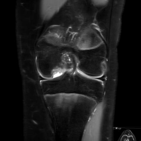
In the MRI examination, DP Dixon in axial, coronal and sagittal sequences were performed. The curved hypodensity correlates with a fracture line extending through the cartilage and subchondral endplate with surrounding subchondral cysts. In the anterior part, the fluid sequence depicts a low signal rim with surrounding oedema of the lesion. In the posterior area, there is a subchondral fluid- filled defect. In the suprapatellar space, there is a free body, with bone and cartilage content.
After the diagnoses, surgery was performed and radiological findings were confirmed.
Discussion
Recently, there have been updates about the terminology of the osteochondral lesions of the knee. Nowadays, the term of Osteochondritis Dissecans (OCD) is the most widely used for an idiopathic fracture line extending through the articular cartilage and the underlying bone in the knee [1, 2]. Within the risk factors, repetitive trauma is accepted [1, 3].
OCD generally affects children and young people [1, 2, 4]. These patients complain about knee pain and swelling, with restricted motion in severe cases with displaced fragment bones [1, 3, 4]. In long-term follow up, the subsequent irregularity of the articular surface can lead to osteoarthritis [1, 3, 4].
CT and X-ray depict a lucent curved fracture in the articular surface, normally affecting the lateral aspect of the medial femoral condyle. In displaced cases, the lucency is focal and accompanied by an intraarticular body with variable bone maturation [1, 2].
MRI is more accurate for OCD diagnosis and can also exclude other chondral damage [1, 2]. It shows the fracture line in the cartilage and the subarticular bone endplate, with adjacent bone oedema in acute cases. In the displaced cases, we can see the bone fragment with variable bone and cartilage components.
Even if the accuracy of the lesion stability is in debate [2, 3, 4], some radiologic signs can help us to distinguish between stable and unstable cases [1, 3]. The instability signs are [1, 2, 3, 4]:
- high T2-signal surrounding the injury, with the same signal intensity of the joint fluid. The signal intensity must be compatible with fluid and must not be confused with granulation tissue [3]
- low T2-signal intensity outer rim, compatible with sclerosis
- multiple or large (more than 5 mm) subchondral cysts
- multiple subchondral breaks
Furthermore, when a displaced fragment is seen, the lesion is also considered unstable. In these patients, we can see the displaced bone fragment with variable bone and cartilage components [1].
The treatment depends on the skeletal maturation and the stability of the lesion [1, 3, 4]. In stable patients, conservative treatment is usually preferred with motion restriction and non-weight-bearing. In unstable or symptomatic cases, drilling, pinning and grafting techniques are performed to encourage vascularisation. Furthermore, excision or fixation of the bone fragments if displacement is done.
Take home message
OCD usually affects the knee in young patients. An early and accurate diagnosis is necessary to prevent long-term consequences.
Written informed patient consent for publication has been obtained.
Differential Diagnosis List
Final Diagnosis
Unstable juvenile OCD with displaced fragment
Liscense
This work is licensed under a Creative Commons Attribution-NonCommercial-ShareAlike 4.0 International License.
Figures


1. Imaging Findings
(1) From the X-ray images (anteroposterior and lateral views), a localized radiolucent area or a sign of separation can be observed in the medial femoral condyle of the left knee (often at the lateral edge of the medial femoral condyle), suggesting discontinuity of the subchondral bone structure.
(2) MRI sequences show a distinct linear signal change involving the cartilage surface and the subchondral bone plate, with a T2 hyperintense band appearing between part of the fragment and the adjacent bone. Localized small fragments or signs of displacement suggest possible osteochondral loosening or separation.
(3) Some sequences reveal bone marrow edema (T2 hyperintensity) and irregularities of the cartilage surface, indicating inflammation or reparative reactions due to repetitive stress or trauma at the lesion site.
(4) Surgical images confirm removal of bone-cartilage fragments, further indicating the presence of detached or highly unstable osteochondral tissue in the corresponding anatomical area.
2. Possible Diagnoses
- Osteochondritis Dissecans (OCD): Considering it typically occurs in adolescents, characterized by knee pain and limited range of motion, with imaging suggesting a linear subchondral bone separation, this is the most likely diagnosis. It commonly occurs in weight-bearing joints, where repetitive microtrauma or impaired blood supply leads to necrosis and loosening of the cartilage and the subchondral bone.
- Osteochondral Injury (Traumatic fracture or cartilage damage): In cases with a clear traumatic event, the possibility of a purely traumatic osteochondral fragment should be considered. However, the clinical presentation is more acute, and the fragment characteristics during surgery may differ from typical trauma-induced fractures.
- Other Rare Intra-Articular Lesions (e.g., Synovial chondromatosis or neoplastic lesions): These usually present with multiple calcifications or mass formation on imaging, which is inconsistent with this case.
3. Final Diagnosis
Based on the patient’s age, chronic knee pain with recent exacerbation, lack of significant history of joint inflammation, and the imaging and intraoperative findings: The most probable diagnosis is Osteochondritis Dissecans (OCD) of the medial femoral condyle, in an unstable state.
4. Treatment and Rehabilitation Plan
Treatment Options:
(1) Conservative Management: For mild and stable lesions, consider reducing weight-bearing activities, using braces or crutches to lessen pressure, and performing physical therapy to facilitate recovery.
(2) Arthroscopic Surgery: In cases of marked instability or presence of loose fragments, arthroscopic evaluation is required to remove the debris, perform drilling, bone-cartilage pinning, or fixation to restore blood supply. If larger fragments remain viable, an attempt can be made to reposition and stabilize them. Where the cartilage surface is severely damaged or fragments are irreparable, cartilage repair procedures or bone grafting may be necessary.
Rehabilitation and Exercise Prescription:
(1) Early Stage (Postoperative Weeks 1–4):
• Primarily immobilize the joint or allow partial weight-bearing. Begin non-weight-bearing isometric quadriceps exercises to maintain muscle strength.
• Incorporate passive or assisted active range of motion exercises to prevent joint stiffness, adjusting speed and range individually.
• Low-intensity training focusing on patellofemoral joint motion and soft tissue mobilization.
(2) Intermediate Stage (Postoperative Weeks 4–8):
• Upon medical clearance, gradually increase joint range of motion and weight-bearing. Stationary cycling and closed-chain exercises (e.g., seated leg press) can help build quadriceps and hamstring endurance.
• Recommended frequency is 3–4 times per week, each session lasting 20–30 minutes, with intensity adjusted to avoid pain or discomfort.
(3) Late Stage (Postoperative Weeks 8–12 or longer):
• Progress to balance and proprioceptive training, such as single-leg stances or BOSU balance exercises, to gradually restore coordination and functionality.
• Carefully introduce light jogging or low-impact aerobic exercises, incrementally increasing duration and intensity.
• If the lesion has healed adequately and joint stability is confirmed, the patient may return to regular sports or physical activities under professional guidance.
Throughout the rehabilitation process, closely monitor knee pain and swelling. Should any significant exacerbation occur, adjust the training regimen accordingly. In patients with compromised bone quality or underlying conditions (e.g., rheumatoid arthritis or autoimmune disorders), emphasize bone density improvement and joint protection measures.
5. Disclaimer
This report is a reference analysis based on current imaging and clinical information and does not replace an in-person examination or professional medical assessment. If you have any concerns or if symptoms worsen, please seek medical attention promptly.
Human Doctor Final Diagnosis
Unstable juvenile OCD with displaced fragment