Pacinian corpuscle hyperplasia


Clinical History
A 58-year-old woman who had worked as a store operator presented with recurring episodes of tenderness on the volar aspect of her left hand for the past two years, which she described as a “burning” or “tingling” sensation. She also described the appearance of subcutaneous tender lumps during those episodes, mainly around the metacarpophalangeal joints and proximal fingers, which then resolved. On presentation, her physical examination was unremarkable, and there was no other relevant past medical history.
Imaging Findings
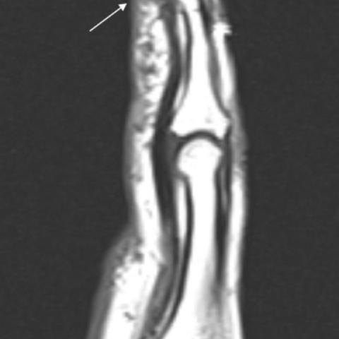
Magnetic resonance (MR) imaging showed multiple millimetric, nodular, round lesions in the subcutaneous plane of the volar surface of the left hand, in greater size and number near the metacarpophalangeal joints and phalanges. The lesions showed very high signal intensity on fluid-sensitive sequences and signal intensity similar to muscle on T1-weighted sequences.
Discussion
Background
Pacinian corpuscles are end-organ mechanoreceptors that respond to changes in pressure and vibration. They are found throughout the body but are most numerous and most tightly grouped in the deep dermis of the volar surface of the hands and feet [1–4]. Normal Pacinian corpuscles are round or ovoid in shape, measure about 1–2 mm in length and are mostly located near the metacarpophalangeal joints and proximal fingers [2–4]. Maximum normal density is 3 to 5 corpuscles per square centimetre [2].
Pathology related to Pacinian corpuscles is rare; there are only a few cases described in the literature, and terminology is inconsistent [2]. “Pacinian corpuscle hyperplasia” refers to an abnormal increase in and/or density of Pacinian corpuscles [1]. The pathogenesis of this condition remains unclear but prior repetitive trauma has been implicated as a potential cause [2].
Clinical Perspective
Pacinian corpuscle hyperplasia predominantly affects women with a mean age of presentation of 49.5 years [1]. It often presents as palpable, subcutaneous tender nodules on the volar aspect of the hands, near the metacarpophalangeal joints and proximal fingers [1,2]. Pain, tenderness, mass sensation, swelling and sensory changes are possible symptoms [1]. Nodules and associated symptoms may spontaneously resolve and later reappear [1,2].
Imaging Perspective
On MR imaging, an increase in the size and number of Pacinian corpuscles is suggestive of Pacinian corpuscles hyperplasia. They appear as multiple nodular lesions on the subcutaneous plane of the volar aspect of the palm and fingers, which show high signal intensity on fluid-sensitive sequences and are isointense to skeletal muscle on T1-weighted sequences. Post-gadolinium administration enhancement is variable [1]. On US, multiple round hypoechoic lesions in the subcutaneous plane may be seen [5]. Although histopathological examination remains necessary to establish a definitive diagnosis [1], typical MR and US findings are very suggestive of this condition in the appropriate clinical setting.
Outcome
Surgical excision is the mainstay of treatment and is curative in the majority of cases [2]. In our case, management consisted of analgesia and clinical follow-up.
Take Home Message / Teaching Points
Pacinian corpuscle hyperplasia is a rare condition that should be considered in the differential diagnosis of tender subcutaneous nodules on the volar aspect of the digits or distal palms. MR and US findings are complementary and are very suggestive of this condition in the appropriate clinical scenario.
All patient data have been completely anonymised throughout the entire manuscript and related files.
Differential Diagnosis List
Final Diagnosis
Pacinian corpuscle hyperplasia
Liscense
This work is licensed under a Creative Commons Attribution-NonCommercial-ShareAlike 4.0 International License.
Figures


Medical Imaging Analysis Report
I. Radiological Findings
Based on the provided MRI images (coronal and sagittal views), multiple small nodular lesions are visible in the subcutaneous tissue on the palmar side of the left hand. These lesions are primarily distributed near the palmar side of the metacarpophalangeal joints and in the deep palmar region of the proximal phalanges. On T2 and other fluid-sensitive sequences, the lesions show relatively high signal intensity; on T1-weighted sequences, the signal is similar to that of muscle. Some lesions display mild to moderate enhancement after contrast administration. The boundaries of the lesions are relatively well-defined, and there are no apparent erosive changes or bony destruction in the surrounding soft tissues or bones.
II. Potential Diagnoses
Considering the patient’s history (periodic burning and stinging pain in the palmar side of the hand, along with palpable subcutaneous nodules), the imaging findings (multiple cystic-like nodular lesions located subcutaneously in the palmar side), and relevant literature, possible diagnoses or differentials include:
- Pacinian corpuscle hyperplasia
Characteristics: Associated with repeated mechanical stimulation or minor trauma, commonly seen in females around 50 years old. Patients may present with periodic pain, burning sensations, or electric shock-like feelings in the palm. Imaging can show multiple small, high-signal nodules, and pathological examination confirms hyperplasia of Pacinian corpuscles. - Giant cell tumor of the tendon sheath
Characteristics: Commonly appears as a solid nodule on the palmar side of the fingers or near joints. On MRI, it often shows low to intermediate signal intensity with possible enhancement, but it is usually solitary. This does not fully match the multifocal pattern seen in this case. - Rheumatoid nodules
Characteristics: Can occur near joints, but typically accompany other clinical symptoms of rheumatoid arthritis, such as morning stiffness and serological abnormalities. No related symptoms or abnormal tests have been reported in this case.
III. Final Diagnosis
Taking into account the patient’s age, symptom characteristics (burning and sharp pain, nodules that may subside and recur), the multiple high-signal nodules on imaging, and literature descriptions of Pacinian corpuscle hyperplasia, the most likely diagnosis is:
Pacinian corpuscle hyperplasia.
For further confirmation, surgical sampling or excision of a larger nodule for pathological examination can be considered.
IV. Treatment Plan and Rehabilitation
1. Treatment Strategies:
• Conservative Management: For patients without marked functional impairment, analgesics (e.g., NSAIDs) or local heat therapy can be used for symptomatic relief.
• Surgical Intervention: In cases of recurrent, significant pain or functional disruption, surgical excision of the most symptomatic nodules can be considered, generally offering good relief.
• Other Measures: If there is any suspicion of trauma or chronic mechanical stress, efforts should be made to minimize excessive or repetitive pressure on the hand.
2. Rehabilitation/Exercise Prescription (FITT-VP Principle):
(1) Frequency: Perform exercises targeting hand flexibility and grip strength 3–4 times per week.
(2) Intensity: Start with low to moderate intensity (e.g., using a grip ball or resistance band where you feel tension but not significant pain).
(3) Time: 15–20 minutes each session initially, gradually increasing to 30 minutes based on tolerance.
(4) Type:
- Finger flexion and extension, making and releasing a fist, possibly with light resistance.
- Wrist flexion, extension, and forearm rotation exercises, progressively increasing the number of repetitions.
- Flexibility routines: passive or active stretching of finger joints, avoiding excessive pain.
(6) Volume: Aim for a total of approximately 90–150 minutes of targeted training per week, based on the patient’s condition.
During the exercises, if pain intensifies, swelling worsens, or other discomfort occurs, training should be stopped immediately and medical evaluation sought. Given the patient’s age, applying heat therapy or localized physiotherapy at the initial stage may help alleviate discomfort and promote circulation.
Disclaimer
This report is a reference analysis based on the current information and imaging findings and does not replace an in-person consultation or professional medical advice. If you have any concerns or notice changes in your symptoms, please consult a qualified healthcare provider promptly.
Human Doctor Final Diagnosis
Pacinian corpuscle hyperplasia