SAPHO syndrome


Clinical History
A 51-year-old female presented with long-standing right upper abdominal pain, minimal weight loss (3 kg in 6 months), and slight night sweats. Laparoscopic cholecystectomy had not resolved the complaints. Except for diffuse hepatic steatosis, abdominal ultrasound was unremarkable. Apart from slightly elevated fibrinogen and liver enzymes, blood tests were normal.
Imaging Findings
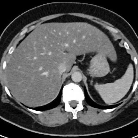
CT thorax and abdomen showed diffuse hepatic steatosis (Fig. 1). The abdominal organs as well as the lungs were otherwise unremarkable, and no lymphadenopathy, pleural effusions, or ascites were noted (Fig. 1). There were diffuse patchy and partly confluent areas of increased osteosclerosis in multiple vertebral bodies of the mid to lower thoracic and the lumbar spine, which were most pronounced at the “vertebral body corners” adjacent to the anterior border of the vertebral endplates (Fig. 2a, b). Sclerosis was as also observed surrounding the left sacroiliac joint, being more pronounced at the iliac side of the joint (Fig. 2c, d). Moreover, there was massive sclerosis of the sternum, mostly affecting the manubrium and extending to the adjoining medial aspects of both clavicles and 1st ribs (Fig. 2a, e–g). The manubrium appeared heavily expanded and there were multiple, bilateral erosive lesions at the costoclavicular joints (Fig. 2a, e).
Discussion
SAPHO (synovitis, acne, pustulosis, hyperostosis, and osteitis) syndrome is a rare chronic inflammatory disorder (estimated prevalence 1/10000 in Caucasians; highest incidence at the age of 30–50; slight female preponderance) of bone, joints, and skin [1–5]. The aetiology is unclear; the pathophysiology is thought to involve genetic, infectious, and immune regulatory factors that lead to an autoinflammatory constellation [6]. Characteristic clinical manifestations are synovitis, osteitis, and hyperostosis, involving the anterior chest wall (65–90% of patients), the sacroiliac joint and spine (32–52%), as well as peripheral joints (<30%) [7–10]. Sacroiliac involvement is typically unilateral and more pronounced at the iliac side of the joint. Cutaneous changes are the second hallmark of SAPHO syndrome, including a variety of acneiform and neutrophilic dermatoses like palmoplantar pustulosis (60% of patients with skin manifestations), nodulocystic acne (25%), or plaque psoriasis [11,12]. Other manifestations or associated pathologies are less frequent, e.g., inflammatory bowel disease [13].
Synovitis results in erosive changes of the affected joints, while osteitis and hyperostosis lead to increased bone density and osseous expansion with cortical thickening and narrowing of the medullary canal [9]. These changes can best be visualised by radiography and CT at later stages of the inflammatory process. MRI can detect active inflammatory changes at much earlier stages, while whole-body bone scintigraphy is useful to detect subclinical lesions with a relatively low inflammatory activity [9]. The “bull’s head sign”, referring to high tracer uptake of the sternocostoclavicular region, is a highly characteristic (but not very sensitive) feature of SAPHO syndrome [14,15].
The patient, in this case, showed many of the typical imaging features including the CT equivalent of the bull’s head sign and sacroiliac joint involvement. Moreover, after raising the suspect of SAPHO syndrome, upon request, the patient reported suffering from relapsing-remitting pustulous and psoriasiform skin changes on both hands and feet (thus far regarded as “psoriasis”, Supplementary Fig. 1-4), as well as upper and lower back pain since her 14th year of life.
Treatment of SAPHO syndrome is mostly empiric since large-scale data are not available. There are various treatment options, including anti-inflammatory and immunomodulatory agents, as well as antibiotics, retinoids, and bisphosphonates [4–6, 11]. The course of SAPHO syndrome can be stable chronic or relapsing-remitting, but disabling complications are rare [4, 5].
This case illustrates the importance of recognising the characteristic imaging features of SAPHO syndrome since its radiologic appearance is key for diagnosis.
Written informed patient consent for publication has been obtained.
Differential Diagnosis List
Final Diagnosis
SAPHO syndrome
Liscense
This work is licensed under a Creative Commons Attribution-NonCommercial-ShareAlike 4.0 International License.
Figures


Supplementary Figure 1

Supplementary Figure 2

Supplementary Figure 3

Supplementary Figure 4

Imaging Findings
1. Liver and Abdomen: From the abdominal CT scans, there are signs of fatty infiltration in the liver, suggesting diffuse fatty liver. No obvious structural abnormalities or masses are seen in the remaining abdominal organs and tissues.
2. Sternum and Anterior Chest Wall: Significant sclerosis and thickening are observed in the sternum and sternoclavicular joint regions. On the coronal CT view, a “bull’s head sign” can be seen — high-density lesions at the manubrium and bilateral sternoclavicular joints, consistent with the chest wall bony sclerosis commonly seen in SAPHO syndrome.
3. Spine: The thoracic and lumbar CT scans show localized bony sclerosis and hyperplasia. There may be focal cortical thickening near the thoracic vertebrae, and parts of the vertebral bodies exhibit increased bone density. However, there is no confirmed pathological fracture or obvious narrowing of intervertebral spaces.
4. Sacroiliac Joint: The CT shows pronounced sclerosis and irregular margins in the unilateral sacroiliac joint, especially on the iliac side. This finding aligns with the typical unilateral sacroiliac joint involvement seen in SAPHO syndrome.
5. Skin: The provided photographs of the palms and feet show recurrent scaling, peeling, pustulous lesions, and some erythema-like changes. These suggest inflammatory skin changes in the hands and feet, featuring psoriasiform or pustular lesions.
Potential Diagnoses
Based on the above imaging findings and clinical presentation, the following diagnoses or differential diagnoses are considered:
1. SAPHO syndrome (Synovitis, Acne, Pustulosis, Hyperostosis, Osteitis): Typical manifestations include sclerotic or hyperplastic lesions of the anterior chest wall, unilateral sacroiliac joint involvement, and pustular skin lesions on hands and feet, matching the patient’s multifocal skeletal and skin findings.
2. Psoriatic Arthritis: Can present with skin and nail changes, as well as joint sclerosis or erosions. However, the “bull’s head sign” feature of the sternum and clavicles is not typical of psoriatic arthritis.
3. Ankylosing Spondylitis: May involve the sacroiliac joints, but usually presents bilaterally, and marked sclerosis or thickening of the sternoclavicular joints is less common.
4. Pyogenic Osteomyelitis or Chronic Infection: Could present with local bony sclerosis, but usually accompanied by overt signs of infection (e.g., local soft tissue swelling, abscess formation) and more severe clinical symptoms. Association with pustular skin lesions is less likely.
Final Diagnosis
Considering the patient’s long-standing recurrent pustular and psoriasiform skin lesions (involving both hands and feet), the characteristic bony thickening and sclerosis of the anterior chest wall and sacroiliac joints (including the “bull’s head sign” visible on CT), and the onset in middle age (around 50 years old) with a chronic, gradually progressive course, the most likely diagnosis is: SAPHO syndrome.
If uncertainty remains, a whole-body bone scintigraphy (to assess for additional lesions) and further dermatological examination or musculoskeletal MRI might be considered to complete the evaluation. However, based on current clinical and imaging features, SAPHO syndrome is highly probable.
Treatment Plan and Rehabilitation
1. Treatment Strategies:
- Non-Steroidal Anti-Inflammatory Drugs (NSAIDs): Can help alleviate joint and bone pain and reduce inflammation.
- Immunomodulators/DMARDs (e.g., methotrexate, sulfasalazine): Suitable for patients with significant chronic inflammatory activity to help control joint and bone inflammation.
- Biological Agents (e.g., TNF-α inhibitors or IL inhibitors): May be considered for patients with recurrent flares or those unresponsive to conventional treatment.
- Topical Skin Treatment: Topical corticosteroids, vitamin A derivatives, or phototherapy can help alleviate hand and foot lesions.
- Antibiotics or Bisphosphonates: Some studies suggest trying these during active bony disease phases, but they should be used based on specific clinical conditions and standardized indications.
- Other Supportive Treatments: Includes correcting or controlling metabolic abnormalities, appropriate nutritional management (e.g., addressing fatty liver), and adjusting the treatment plan as needed based on disease activity.
2. Rehabilitation/Exercise Prescription:
Given that SAPHO syndrome often involves bony hyperplasia and joint involvement, particular attention should be paid to the flexibility and stability of the spine and sacroiliac joints. Recommended approach:
- Initial Phase (Weeks 1–2):
· Focus on low-intensity joint mobility and stretching exercises (e.g., small pelvic rotations or leg raises in a supine or seated position).
· Perform 10–15 minutes per session, 3–4 times per week, avoiding vigorous joint impact and twisting.
- Progression Phase (Weeks 3–6):
· Increase core and back muscle strength training using moderate resistance such as elastic bands or light dumbbells for stretching and resistance exercises.
· Perform 15–20 minutes per session, 3–5 times per week, maintaining moderate-to-low intensity and monitoring joint and bone responses.
- Stabilization Phase (After 6 Weeks):
· If symptoms improve, engage in low-impact aerobic activities (e.g., swimming, brisk walking, or cycling) after adequate warm-up, for 20–30 minutes per session, 3–5 times per week.
· Incorporate light-weight resistance training (dumbbells, resistance bands) to strengthen core and iliopsoas muscles, avoiding high-impact activities on the sternum, clavicles, and sacroiliac joints.
- Individual Adjustments: Modify the regimen according to the patient’s pain, fatigue levels, and skin condition. If symptoms noticeably worsen or discomfort arises, prompt re-evaluation is advised.
Safety Notice: Due to bony changes and chronic pain in this condition, ensure adequate warm-up and gradual adjustment of exercise intensity. When necessary, collaborate with a specialized rehabilitation therapist or sports medicine specialist to develop an individualized exercise program.
Disclaimer: This report is a reference analysis based on the available information and does not replace an in-person consultation or professional medical opinion. For further questions, please consult a specialist or seek medical attention.
Human Doctor Final Diagnosis
SAPHO syndrome