Morel-Lavallée lesion - a reminder of a small piece in the trauma puzzle


Clinical History
A 44-year old patient was referred to the radiology department to undergo a thoracic, abdominal, and pelvic CT scan after suffering a fall from height (~ 2 meters). The CT scan revealed a soft-tissue lesion located superficially at the right upper thigh area, which was furthermore investigated by ultrasound and MRI.
Imaging Findings
Various imaging techniques can be used to diagnose a posttraumatic lesion. In our case, CT was the first to raise suspicion for the diagnosis, which was confirmed by ultrasound and MRI.
CT shows a high-density collection with heterogeneous structure, including fat, liquid, and blood degradation products, associated with edematous infiltration of surrounding adipose tissue. CT confirms the lesion's superficial location in the interfascial plane, between hypodermis and deep fascia.
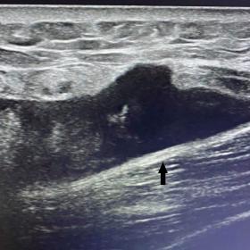
Ultrasound demonstrates a heterogeneous hypoechoic lesion located in the subdermal plane immediately above the muscular fascia, with some internal fat echoes; lack of vascularity on colour Doppler examination is an important feature.
MRI's role in the final diagnosis is essential by demonstrating the lesion's topography and internal structure. T1 FS sequences show a hyperintense lenticular collection, with some internal fat globules and thin septa. T1 trim sequence emphasizes the adjacent inflammatory alterations in the hypodermis and the collection's serious component. T2* sequence reveals the presence of blood degradation products.
Discussion
The Morel-Lavallée lesion (MLL) is a post-traumatic closed degloving injury where the subcutaneous tissue is traumatically separated from the underlying deep fascial layer, creating a potential space that is progressively occupied by blood, lymph, and/or liquified fat. [1,2]
MLL is an uncommon lesion. The literature describes an 8.3% prevalence in the context of pelvic trauma, with a 2:1 male to female ratio. [3]
MLL may appear as a result of blunt force trauma or crush injuries where high‐intensity shearing forces are applied tangential to the fascial plane. Most frequently it involves the peritrochanteric region and the proximal thigh. [3]
Clinically there is often a painful focal area of swelling. Physical examination reveals a compressible, fluctuant lesion. [4]
Diagnosis is based on medical history, clinical examination, and imaging techniques, including ultrasound, CT, and MRI. MRI is the gold standard for the description and diagnosis of MLL. [5]
Ultrasound is a rapid and non-expensive method, but highly non-specific, demonstrating a variable appearance of MLL, depending on the age of the lesion. It often shows a heterogeneous hypoechoic collection with intralesional septations and/or hyperechoic fat globules, or rarely, a homogeneous anechoic lesion. The most important features demonstrated by the ultrasound examination are the absence of internal vascular flow and the lesion's location superficial to the muscle fascia and deep to the hypodermis. Sometimes a chronic lesion may be surrounded by a vascularized capsule. [2,6]
CT usually is the initial modality of investigation in acute trauma cases. It depicts the presence of a fluid collection (relatively high density, within 15-40 Hounsfield units, but sometimes with various internal densities, like that of fluid, fat, or blood), with the typical localization. In an acute presentation, MLL is ill-defined, with surrounding fat-stranding, while chronic lesions are encapsulated. [1,3]
The MRI appearance varies depending on the content (the concentration of hemolymphatic fluid) and chronicity of the lesion: homogenous signal intensity and smooth margins (sometimes an enhancing capsule) if chronic, or heterogeneous signal and irregular limits, with surrounding soft-tissue oedema if acute. These characteristics are reunited in the Mellado-Bencardino classification, which describes the appearance on T1 and T2-WI, as well as other features like the shape and enhancement characteristics. The demonstration of adipose signal intensity within the lesion, and also GRE/T2* sequences, which are useful for revealing the internal presence of blood degradation products through the blooming artefact are valuable clues towards the diagnosis. [1,3,7]
The treatment is different depending on the chronicity of the lesion and the presence of associated conditions (superinfections, bone fractures). Acute MLL is conservatively treated with compression, while chronic lesions are reserved for percutaneous aspiration and sclerotherapy. Complicated cases (superinfected, with skin necrosis), associated with a late diagnosis are surgically addressed. [3,5]
In conclusion, MLL is an uncommon posttraumatic lesion with an imaging-based diagnosis, which radiologists should be aware of because early diagnosis permits an easier, conservative treatment.
Written informed patient consent for publication has been obtained.
Differential Diagnosis List
Final Diagnosis
Morel-Lavallée lesion.
Liscense
This work is licensed under a Creative Commons Attribution-NonCommercial-ShareAlike 4.0 International License.
Figures
Ultrasound caption (1a), Color Doppler Ultrasound (1b)


Axial (2a) and coronal (2b) non-contrast CT


T1 FS axial (3a), T1 tirm coronal (3b)


T2*coronal

1. Imaging Findings
Based on the provided ultrasound, CT, and MRI images, there is a noticeable fluid-like lesion in the superficial soft tissue region of the right upper thigh. This lesion lies between the subcutaneous fat and the deep fascia, appearing irregular or oval. At different stages, varying degrees of internal density changes or mixed signals can be observed.
- Ultrasound: The lesion appears as a relatively hypoechoic or mixed echo area, sometimes with septations or internal fatty-density echoes, and a relatively clear margin in certain regions. No significant blood flow signals are detected inside.
- CT: A collection within the soft tissue shows a density slightly higher than pure fluid (often in the 15-40 HU range), with some areas displaying fat density or hemorrhagic components. In the acute phase, it may appear loose with irregular borders, indicating peripheral tissue exudation or infiltration. Over time, a capsule could form.
- MRI: On T1WI and T2WI, the lesion’s signals can vary due to the mixture of blood and lymph. In chronic stages, the margins can be relatively well-defined with an enhancing capsule; newly formed lesions may present with heterogeneous signals and surrounding soft tissue edema.
2. Potential Diagnoses
- Morel-Lavallée Lesion: Commonly seen in trauma that separates the subcutaneous fat layer from the deep fascia, creating a potential fluid collection space containing blood, lymph, and liquefied fat. Imaging typically shows a large fluid collection between the subcutaneous and fascial layers with mixed density or signal, sometimes indicating old hemorrhage or fat.
- Subcutaneous Hematoma: Post-traumatic local hematoma can also appear as a fluid collection, especially in the acute stage, with uneven echoes on ultrasound or higher density on CT. However, most subcutaneous hematomas are confined to the dermal layer or superficial fat, making significant separation between the fascia less common.
- Soft Tissue Fluid Collection or Cyst: Such as a serous cyst or fat-liquefied cyst in the subcutaneous tissue, generally lacking traumatic history and rarely showing mixed signals or capsule enhancement on MRI or CT.
Considering the clinical history of trauma (fall from a height) and the imaging findings (lesion located between subcutaneous and deep fascial layers with mixed echogenicity/density/signals), a Morel-Lavallée lesion is highly suspected.
3. Final Diagnosis
Given the clear traumatic history and imaging demonstrating fluid accumulation between the subcutaneous and deep fascial layers, along with characteristic MRI and CT findings, the most likely final diagnosis is a Morel-Lavallée lesion.
If any doubt remains, aspiration or biopsy during treatment may be performed for pathological examination to rule out rare infections or neoplasms.
4. Treatment Plan and Rehabilitation
4.1 Treatment Strategy
- Conservative Treatment in the Acute Phase: Early use of compression bandaging or elastic dressings helps reduce subcutaneous fluid exudation and prevent recurrent accumulation.
- Intervention in the Chronic Phase: For encapsulated lesions or those with large fluid volume, aspiration combined with sclerotherapy can be considered. If there is skin necrosis, infection, or frequent recurrence, surgical debridement and capsule excision may be needed.
- Management of Complications: In cases of associated fractures, infections, or skin ischemic necrosis, address these primary problems concurrently. Post-healing, appropriate rehabilitation should be conducted.
4.2 Rehabilitation Exercises and Exercise Prescription
During soft tissue repair after trauma and in postoperative recovery, a gradual exercise program can improve circulation, reduce edema, and maintain muscle function. Rehabilitation recommendations are as follows:
- Early Stage (Acute/Subacute):
- Reduced Weight Bearing: Avoid excessive activity of the affected side, possibly using braces or crutches.
- Isometric Muscle Exercises: Perform isometric contractions of the quadriceps as tolerated, avoiding tension that stretches the subcutaneous space.
- Each session lasts 10-15 minutes, 1-2 times per day.
- Middle Stage (Stable Recovery):
- Gentle Joint Movements: Perform passive and active exercises of the hip and knee within a tolerable pain range.
- Low-Resistance Training: Incorporate gentle resistance exercises with elastic bands to build strength.
- Each session lasts 15-20 minutes, 2-3 times a week, with gradual increases in duration and intensity as tolerated.
- Late Stage (Return to Function):
- Progressive Functional Training: Include jogging, cycling, and core exercises, avoiding direct impact to the affected area.
- Strength and Flexibility Work: Gradually enhance lower limb strength and soft tissue flexibility to reduce adhesions and prevent re-injury.
- Each session lasts about 30 minutes, 3-4 times a week, and can be gradually increased to normal training levels.
Throughout the rehabilitation process, an individualized approach following the FITT-VP Principle (Frequency, Intensity, Time, Type, Volume, Progression) should be employed. Monitor the wound and local tissues closely during activities, and consult with a physician or rehabilitation specialist as needed.
Disclaimer: This report is a reference-based analysis and cannot replace face-to-face consultation or professional medical judgment. If you have any questions or if symptoms worsen, please seek prompt medical attention.
Human Doctor Final Diagnosis
Morel-Lavallée lesion.