A teenager with hip pain


Clinical History
A teenager presented with a 10 months history of worsening left hip pain, aching at night and limping. On examination there was muscle wasting and restriction to hip movements. Inflammatory markers including CRP and full count were normal. The pain improved with anti-inflammatory medication. No other joints were affected.
Imaging Findings
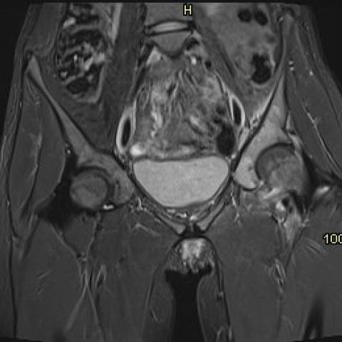
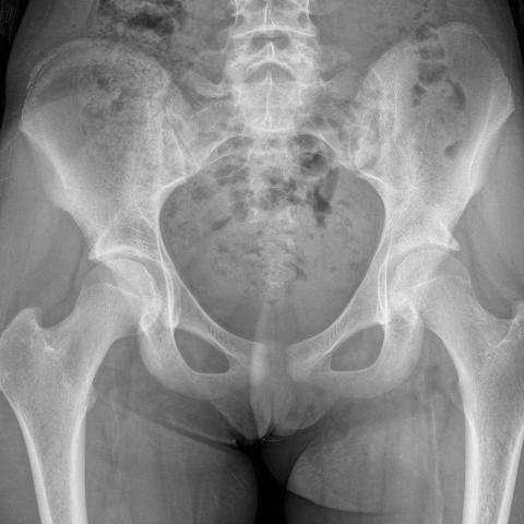
Marked marrow oedema is seen on fluid sensitive Magnetic Resonance (MR) sequences in the left acetabulum extending into the left ilium (Fig. 1a). There is a small left hip joint effusion (Fig. 1b). Post gadolinium there is enhancement in the acetabulum, the synovium of the hip joint and marked enhancement in the soft tissues anterior to the hip surrounding the psoas tendon (Fig. 1c). The nidus at the anterior acetabular rim is seen on thin-section axial T2 fat-saturated MR images as a high signal rim surrounding a low signal centre (Fig. 2). Sclerosis surrounding the nidus is best seen on Computed tomography (CT) (Fig. 3) but may be difficult to appreciate on plain radiographs (Fig. 4). Single Photon Emission Computed Tomography (SPECT) shows a focus of marked increased tracer uptake in the nidus at the anterior acetabulum (Fig. 5).
Discussion
Background
Osteoid Osteoma is a benign primary bone tumour more common in young males [1, 2]. It is typically seen in the cortex of long bones with predilection for the lower extremity. The skull, scapula, ribs and pelvis are rare locations. The nidus of the tumour secrets prostaglandins leading to classic symptoms of night pain promptly relieved by Salicylates [3].
Clinical perspective
Intraarticular osteoid osteoma, a lesion occurring near a joint is most common in the hip. Often regarded as a different clinical entity, symptoms are non-specific with arthralgia and joint stiffness [1]. Patients may not have night pain or joint pain relieved by salicylates [1]. Joint effusions may be attributed to other causes delaying the diagnosis. Treatment commonly comprises surgery, radiofrequency ablation, CT-guided drill resection or conservative treatment.
Imaging perspective
On MR imaging the nidus shows low to intermediate signal intensity on T1 weighted sequences and imaging characteristics on T2 weighted sequences change depending on the amount of central mineralisation. It typically shows a peripheral rim of high signal on T2 weighted sequences with a low signal centre [4]. Post gadolinium, the nidus may show enhancement. Earlies studies showed that compared with CT, MR imaging may fail to demonstrate a small nidus [4, 5]. However, recently Zampa et al. suggest that dynamic contrast enhanced MR imaging increases nidus visibility and helps to identify lesions in atypical locations [4, 6].
On CT the nidus is depicted as a round or oval low attenuation lesion with an area of central high attenuation that represents mineralised osteoid surrounded by reactive sclerosis.
This sclerosis may highlight a small nidus on radiographs. However, the amount of sclerosis can be minimal for intraarticular lesions, especially in the hip, or only appreciated retrospectively on radiographs. Therefore, the nidus may be not be appreciated [1]. This is because the intraarticular periosteum cannot generate a strong sclerotic periosteal response as caused by lesions in an extracapsular location [2]. Periosteal reaction can be seen outside the joint [1].
Intraarticular osteoid osteoma can be associated with excessive surrounding marrow oedema, soft-tissue oedema, synovial hypertrophy and joint effusion making an accurate diagnosis more difficult [7]. In select cases this may also prompt biopsy to verify the final diagnosis.
On technetium-99m methylene diphosphonate bone scans the nidus shows intense uptake which can aid diagnosis for lesions in atypical locations [1].
Teaching points
- Intraarticular osteoid osteomas often have atypical clinical presentations.
- Severe associated inflammatory change may distract from the diagnosis.
- Sclerosis surrounding intraarticular osteoid osteoma can be minimal but periosteal reaction may be seen outside the joint capsule.
Written informed patient consent for publication has been obtained.
Differential Diagnosis List
Final Diagnosis
Acetabular, intra-articular osteoid osteoma
Liscense
This work is licensed under a Creative Commons Attribution-NonCommercial-ShareAlike 4.0 International License.
Figures


Imaging Findings
Based on the provided MRI, CT, and X-ray images, the lesion is located near the left hip joint (adjacent to the proximal femur). MRI shows varying degrees of edema signals in the soft tissues around the joint, with mild joint effusion and synovial reaction visible in the local joint space. A possible low- to medium-signal core is observed in the lesion area (more prominent on T1-weighted images), surrounded by a high-signal ring structure on T2-weighted images, suggesting features of an active lesion. On the CT images, a round or oval low-density zone is seen in the cortex of the left proximal femur, with a center that appears as a high-density calcification-like area. Surrounding sclerosis is not prominent, although mild bone sclerosis and reactive changes in the local bone cortex are noted. On routine X-ray, the lesion is not clearly visible due to the lack of significant reactive bone sclerosis; thus, CT and MRI are necessary for better localization of the lesion.
Potential Diagnoses
- Intra-articular Osteoid Osteoma
Typical presentations include nocturnal pain relieved by nonsteroidal anti-inflammatory drugs (NSAIDs). The lesion often manifests as a small round/oval “nidus” with associated bone sclerosis. Because this case is near a joint, sclerosis may be mild, and MRI often demonstrates pronounced soft tissue or bone marrow edema.
- Other Benign Bone Tumors (e.g., Osteochondroma)
On imaging, these tumors often show other types of calcifications or a cartilage cap, with varying degrees of periosteal changes. However, they may not fully match the typical nocturnal pain that is rapidly relieved by NSAIDs.
- Occult or Subacute Osteomyelitis
In patients with a history of chronic bone pain, infection should be considered. However, in this case, inflammatory markers (CRP, complete blood count, etc.) are normal, and the pain is relieved by NSAIDs, making infection less likely.
- Osteoarthritis or Juvenile Idiopathic Arthritis
Although these conditions can cause joint pain and restricted movement, they are commonly associated with complex autoimmune or degenerative factors. Nocturnal pain rapidly relieved by NSAIDs is not typically characteristic.
Final Diagnosis
Taking into account the patient’s age (14 years old), the characteristic nocturnal pain and significant relief by NSAIDs, along with imaging findings (an apparent “nidus” lesion on MRI and mild sclerosis with a central calcification-like area on CT), the most likely diagnosis is intra-articular osteoid osteoma (left hip area, proximal femur). In this location, osteoid osteomas often present with minimal surrounding sclerosis while displaying joint effusion, soft tissue swelling, and other inflammatory signs. If uncertainty remains, an image-guided biopsy of the lesion can be considered for further confirmation.
Treatment Plan and Rehabilitation
- Treatment Strategies
- Conservative Treatment: If symptoms are tolerable, continue using NSAIDs for pain control and observe. If symptoms steadily improve and the lesion does not enlarge, regular outpatient follow-up can be maintained.
- Minimally Invasive Treatment: CT-guided radiofrequency ablation (RFA) or drilling excision can quickly relieve pain and remove the lesion with relatively minor trauma. This is suitable for patients with marked symptoms, poor response to conservative treatment, or when a definitive diagnosis is established.
- Surgical Treatment: If imaging makes RFA targeting difficult, or if there are indications for surgery (such as a larger lesion, suspected malignancy, or impaired joint function), surgical resection may be considered. However, the extent of damage and impact on joint stability must be evaluated.
- Rehabilitation and Exercise Prescription (FITT-VP Principle)
- Frequency (F): Perform functional exercises 3–4 times per week.
- Intensity (I): Begin with low-intensity joint activities such as swimming and light cycling. As pain is controlled, intensity can be gradually increased.
- Time (T): Each session should last about 20–30 minutes. If fatigue or pain worsens significantly, reduce the duration.
- Type (T): Emphasize non-weight-bearing or low-weight-bearing exercises, such as seated cycling, water activities, or wall squats (with shallow range). Progressively incorporate flexibility and muscle-strengthening exercises for the joint.
- Progression (P): Once symptoms improve, slowly introduce weight-bearing exercises and core strengthening, for example, short-step jogging and resistance band training for the lower limbs. Regularly assess hip joint function and pain level, adjusting weight-bearing and exercise volume accordingly.
- Key Considerations: If significant pain, joint swelling, or locking sensations occur, stop the activity and consult a doctor for evaluation. In adolescents with low bone density or inadequate fitness, consider appropriate nutritional support and rest, ensuring a gradual, safe progression.
Disclaimer:
This report is a reference analysis based on existing images and data and cannot substitute an in-person consultation or advice from a qualified physician. Specific treatment and rehabilitation should be tailored to the patient's individual situation and guided by specialist evaluation.
Human Doctor Final Diagnosis
Acetabular, intra-articular osteoid osteoma