Child with forefoot pain and swelling of insidious onset


Clinical History
A 7-year-old boy, Tae-Kwon-Do athlete, presented with insidious onset of pain and swelling of the right foot over two months, without any history of trauma. Pain initially presented during sports activities but gradually it became constant during everyday activities. Clinical examination revealed tenderness over the first metatarsal. Medical history was unremarkable.
Imaging Findings
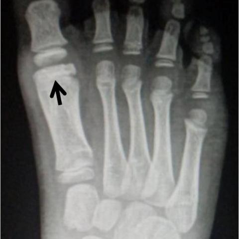
Anteroposterior and oblique plain foot radiographs demonstrated flattening, sclerosis, without significant fragmentation of the distal first metatarsal epiphysis (Fig. 1). MR with STIR and T2-weighted (T2W) images confirmed articular collapse and necrosis of the distal epiphysis which appeared with low signal intensity surrounded by extensive high signal bone marrow oedema of the metatarsal diaphysis (Fig. 2). A subchondral linear fracture was also shown as a linear low signal intensity lesion within the bone marrow oedema.
Discussion
Background
Freiberg’s disease (FD) is an idiopathic avascular necrosis located in the second metatarsal head in 2/3 of cases and the third in about 1/3 [1]. Involvement of the lesser toes and first metatarsal is extremely rare [1-3]. The pathophysiology of FD is still unclear. Vascular damage due to repetitive microtrauma and fractures of the subchondral bone may be the underlying cause [4]. FD has been linked to high-heeled shoes and its prevalence is higher in women at their second decade of life [5,6].
Clinical Perspective
Clinical examination reveals swelling and tenderness of the affected metatarsophalangeal joint. History may or may not reveal previous trauma. Disease progression leads to foot deformity with a claw or crossover toe appearance. Conservative treatment is the mainline of management, along with non-steroidal anti-inflammatory medication, activity modification and use of rocker bottom shoes. Surgical treatment is reserved only for cases of advanced disease or when conservative treatment has failed and includes a combination of debridement, loose body removal, osteotomies and core decompression [4,5]. In our case, repetitive microtrauma due to intensive Tae-Kwon-Do training was postulated to be the causative factor. Despite the atypical age, sex and location, imaging findings and clinical history were highly specific for FD.
Imaging Perspective
Plain films may demonstrate sclerosis, flattening and fragmentation of the affected metatarsal head. MRI is the imaging modality of choice since it can detect the disease at early stages when findings in plain films are absent or subtle [7]. At the initial stages, MRI shows low signal intensity of the metatarsal head on T1W and high signal intensity on fluid sensitive sequences. As necrosis evolves, the metatarsal head appears with low signal intensity on all pulse sequences. Bone marrow oedema of the surrounding bone, subchondral fracture lines and metatarsophalangeal joint effusion may also be present [1,6,7]. The disease can be classified to five stages of severity according to Smillie, which works both for plain films and MRI [5]. Surgical treatment is reserved for advanced disease.
Outcome
Conservative treatment usually provides full resolution of symptoms and imaging findings. In our case, due to the demand for return to high level of performance, the patient was treated surgically with debridement and core decompression. Complete resolution of symptoms and imaging findings were noted at the 1.5 year follow-up (Fig. 3).
Written informed patient consent for publication has been obtained.
Differential Diagnosis List
Final Diagnosis
Freiberg’s disease of the first metatarsal head in a child
Liscense
This work is licensed under a Creative Commons Attribution-NonCommercial-ShareAlike 4.0 International License.
Figures


Medical Imaging Analysis Report
I. Radiographic Findings
The patient is a 7-year-old boy who has been experiencing pain and swelling in the area of the first metatarsal head on the right foot for about two months. The provided X-ray images show:
- An abnormal shape of the first metatarsal head, with focal sclerosis, flattening, and fragment-like changes.
- A possible slight change in the joint space, though no obvious collapse of the articular surface is noted.
- Mild swelling in the adjacent soft tissue, with no clear fracture lines or free bone fragments observed.
Regarding the MRI findings:
- The first metatarsal head shows low signal intensity on T1-weighted images, consistent with necrosis or altered bone structure.
- T2/PD-weighted and fat-suppression sequences reveal high signal abnormalities, indicating bone marrow edema and potential subchondral fracture lines.
- A small amount of joint effusion is seen around the joint, suggesting reactive changes.
Overall, the imaging findings suggest avascular necrosis of the first metatarsal head, accompanied by fragmented bone structure, sclerosis, and edema signals.
II. Possible Diagnoses
Based on the patient’s age, symptoms, and imaging findings, the possible diagnoses or differential diagnoses include:
- Freiberg disease (avascular necrosis of the metatarsal head): It typically involves the smaller metatarsal heads, but may occasionally occur in the first metatarsal head. Classic findings include metatarsal head collapse, fragmentation, sclerosis, and periarticular edema.
- Other metatarsal osteochondropathies: For example, Köhler disease more commonly affects the navicular bone, but similar osteochondral lesions need to be ruled out.
- Bone or joint infection (e.g., osteomyelitis or septic arthritis): Generally accompanied by obvious fever or an acute exacerbation of pain. Imaging often shows marked bone destruction or soft tissue abscess formation, which is not evident here.
- Benign bone tumors or bone cysts: Such as osteochondroma or simple bone cyst. However, these typically show different imaging features, often including a more localized lesion with osteolytic expansion changes.
Based on the radiographic characteristics and the clinical course, Freiberg disease is strongly considered as the primary diagnosis.
III. Final Diagnosis
Taking into account the patient’s age (even though the gender differs slightly from the typical demographic), regular physical activity (chronic microtrauma possibly from Taekwondo training), and the imaging findings (sclerosis and fragmentation of the first metatarsal head, along with significant bone marrow edema and signal alterations on MRI), the most likely diagnosis is:
Freiberg disease (avascular necrosis of the first metatarsal head).
If uncertainty remains, further clinical evaluations (laboratory inflammatory markers, tests to rule out infection or rheumatic causes) could be performed to exclude other etiologies. However, based on the current data, Freiberg disease is the most probable diagnosis.
IV. Treatment and Rehabilitation Plan
Treatment options can be divided into conservative and surgical approaches, which should be chosen depending on the patient’s activity requirements and the severity of the lesion:
1. Conservative Treatment
- Reduce weight-bearing on the affected foot: Suspend or reduce high-impact activities (such as Taekwondo kicking, running, and jumping) to prevent further injury.
- Medication: Use nonsteroidal anti-inflammatory drugs (NSAIDs) to relieve pain and inflammation.
- Foot orthoses or specialized shoe inserts: Consider using rocker sole shoes or arch supports to offload stress on the metatarsal head.
- Physical therapy: Adjuvant therapies during rehabilitation (e.g., hot compresses, ultrasound, low-intensity shockwave therapy) to improve local blood flow.
2. Surgical Treatment
If the lesion is relatively advanced, or if conservative treatment fails to provide sufficient relief, surgical options include:
- Debridement and core decompression: Remove necrotic and fragmented bone and cartilage, reduce joint pressure, and promote revascularization.
- Osteotomy (if there is significant deformity): Realign the metatarsal’s weight-bearing axis to enhance joint function.
In this case, if the child has higher sporting demands and cannot achieve adequate functional recovery through conservative means, surgical intervention may be considered.
3. Rehabilitation and Exercise Prescription
Since the patient is still young and the skeletal system is in a developmental phase, rehabilitation and exercise prescriptions must be individualized and progressive. The FITT-VP principle may be applied:
- Frequency: Begin with low-intensity activities 1–2 times per week, gradually progressing to 2–3 times per week once pain is under control.
- Intensity: Remain at a level that does not cause significant pain or fatigue. Begin with simple lower-extremity strengthening and flexibility exercises (e.g., seated leg raises, band-assisted exercises).
- Time: Start with 15–20 minutes per session, progressively increasing to 30 minutes based on tolerance. Avoid maintaining a single position for too long.
- Type: Select low-weight-bearing activities such as swimming or cycling to minimize impact on the first metatarsal head; gradually transition to light jogging or lower-intensity Taekwondo foundational drills.
- Volume & Progression: Increase activity volume and intensity as symptoms improve, monitoring foot pain and function closely to avoid overtraining.
In the early stages of rehabilitation, pay close attention to maintaining range-of-motion exercises for the affected foot but avoid high-intensity kicking movements. If significant pain or discomfort arises during training, stop immediately and seek a follow-up evaluation.
Disclaimer: This report is based solely on the current imaging and clinical information available. The opinions provided are for reference only and cannot replace an in-person consultation or a professional physician’s direct diagnosis and treatment plan. If you have any questions or if symptoms worsen, please seek medical attention promptly.
Human Doctor Final Diagnosis
Freiberg’s disease of the first metatarsal head in a child