


A middle-aged female came with complaints of back pain for 3 months. There was no history of trauma, fever or paraesthesia. On clinical examination, there was tenderness in the thoracic region without any kyphosis or scoliosis.
CT and MRI of the dorsal spine showed an osteolytic lesion in the D9 vertebral body with extension to both pedicles, right transverse process, right lamina, right superior articular process and thinning with focal destruction of cortex in right lateral aspect. A large extraosseous enhancing soft tissue component was seen extending through the defect to right paravertebral region with stretched periosteum around the lesion, from D8 to D10 levels with effacement of right lateral recess and neural foramina and indentation of exiting nerve roots. Irregular calcifications were seen in the peripheral part of the extraosseous soft tissue components. Screening of the rest of the axial skeleton was unremarkable.
Background
Giant cell tumour (GCT) of bone is a locally aggressive benign tumour that usually involves the metaphyseoepiphyseal location of long bones. Histologically it is seen as a proliferation of mononuclear stromal cells and homogeneous distribution of multinucleated giant cells [1]. They comprise about 4-9.5 % of all primary bone neoplasms and 18-23 % of all benign bone neoplasms [2]. GCT of the spine is an uncommon occurrence, with the sacrum being the most frequent site comprising 1.7-8.2% of cases and occurrence in mobile spine was only about 2-4% of cases [3]. There is no separate data for cervical, dorsal and lumbar spine, however, there are scattered reports which suggest the rare occurrence of GCT in dorsal vertebra. There is a slight female predilection, especially when located in the spine [2]. Previous studies reveal that any sites other than the sacrum in the spine are a rarer phenomenon still.
Clinical Perspective
Presenting complaints of GCT usually include back pain, soft tissue mass, compression of the spinal cord, and nearby structures. Our patient came with complaints of back pain for 3 months.
Imaging Perspective
Imaging findings suggestive of GCT include an expansile osteolytic lesion producing collapse of the vertebral body and adjacent soft tissue masses. CT shows soft tissue masses without mineralized matrix, sclerotic margins in the eccentric side of the lesion. MR shows predominant T2 hypointensity which could help differentiating from other pathologies which are predominantly T2 hyperintense [4]. Our case showed an osteolytic lesion with sclerotic margins in the eccentric side and showed T2 heterogeneity with post-contrast enhancement.
Outcome
Post imaging the patient underwent CT guided biopsy and HPE revealed giant cell tumour of the dorsal vertebra. The patient underwent en-bloc spondylectomy and thoracic paraspinal tumour excision, cage fixation, and long stabilization of D7 to D11 were done. HPE showed a highly cellular biphasic tumour interspersed with numerous multinucleated osteoclast types of giant cells, consistent with grade II giant cell tumour of the dorsal vertebral body as per classification system proposed by Jaffe et al). [5] Prognostic significance of this histological grading is controversial. It is agreed to some extent that intra-lesional curettage rather than wide resection provides adequate tumour eradication for the majority of patients with GCT. [6] Additional treatment options especially for GCT of the spine involves en bloc resection, preoperative embolization and neoadjuvant radiotherapy and chemotherapy with cisplatin and doxorubicin [7,8].
Take-Home Message / Teaching Points:
We present this case because of the rare occurrence of the GCT of the dorsal spine, a multidisciplinary approach is always needed to diagnose these cases and plan treatment approaches accordingly.
Written informed patient consent for publication has been obtained.
Giant cell tumor of the dorsal vertebral body
This work is licensed under a Creative Commons Attribution-NonCommercial-ShareAlike 4.0 International License.


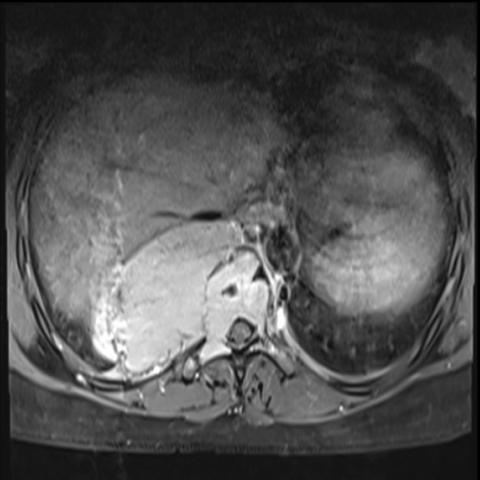

Based on the provided CT and MRI images, there is a localized expansile, osteolytic lesion in the 7th thoracic vertebra (D7). Partial sclerosis is seen around the margins of the lesion. The CT images show a low-density area in the vertebral body with eccentric destruction, and the cortical bone is locally thinned or slightly bulging. On MRI, the affected region shows mixed signals. In T1-weighted images, signals are predominantly low to intermediate, and in T2-weighted images, some regions show relatively low signal along with a certain soft tissue component, consistent with the characteristic “relatively low T2 signal” often seen in some GCTs. No obvious large paravertebral soft tissue mass is observed, but a slight soft tissue bulge is noted around the vertebral margin. Surrounding vertebrae and the posterior elements do not show any significant destruction, and there is no evident scoliosis or kyphotic deformity.
Taking into account the patient’s age (35 years), clinical presentation (persistent back pain for 3 months with no obvious trauma or other systemic symptoms), imaging findings (eccentric osteolytic lesion in D7 vertebra with partial sclerosis and predominantly low T2 signal on MRI), and pathological examination (biopsy confirmed giant cell tumor), the final diagnosis is:
Giant Cell Tumor of Bone (GCT) in the 7th Thoracic Vertebra (D7).
Treatment Strategy:
Rehabilitation and Exercise Prescription: Postoperative rehabilitation should be carried out cautiously to maintain spinal stability and gradually restore motor function.
This report is intended solely for reference based on the available information. It cannot replace in-person consultations or professional medical advice. For actual diagnosis and treatment plans, please consult a specialist physician or orthopedic oncologist who can make final decisions based on the patient’s individual condition and the latest medical guidelines.
Giant cell tumor of the dorsal vertebral body