A case of brucella osteomyelitis of the femur


Clinical History
A 4-year-old male came to a hospital with complaints of undulating fever, femurs pain, impaired motor function in the lower limbs. For two years, the complaints increased and the patient was referred to our hospital with lower limb deformities and with a suspicion of a fracture of the right femoral neck.
Imaging Findings
A plain X-ray of the pelvis and knee joints was performed. It showed shortening of the diaphysis of the right femur, calcification of the soft tissues of the femur on both sides, in the proximal left tibia. Such extensive calcification might be due to the often long, chronic, subclinical course of the disease. The x-ray showed that the iliosacral joints were intact, without any pathological changes. (Fig.1a). It also showed erosion of the condyle of the left femur, preservation of a fairly uniform non-narrowed joint space, and no changes in the condyles of the tibia and patella (Fig.1b).
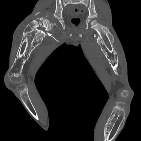
Pelvic computed tomography showed a comminuted fracture of the right femoral neck with a significant periosteal reaction, nonunion of bone fragments, pseudarthrosis of the right femoral neck (Fig.2).
A biopsy of the right femur was performed, a culture of Brucella melitensis was isolated. Antibodies to Brucella melitensis were also detected in blood samples. The patient was diagnosed with chronic brucellosis infection. The management of the patient included surgical treatment: detorsional supracondylar osteotomy of the left femur with plastic defect correction at the level of the distal metaepiphysis, followed by fixation with a cellacast spica cast, and antibiotics in combination therapy.
Discussion
Brucellosis is a particularly dangerous and socially significant zoonotic infection caused by aerobic Gram-negative bacillus of the genus Brucella. Brucella melitensis is considered the most pathogenic species for humans [1]. Endemic areas for brucellosis include countries of the Mediterranean Basin, the Middle East, Asia, the Indian subcontinent, Africa
[2,3]. Brucellosis can affect various organs and tissues: most often it is the osteoarticular system (30-85%) with arthritis, bursitis, sacroiliitis (up to 54%), spondylitis (2-50%), osteomyelitis [4,5,6]. Osteomyelitis is an extremely rare complication of brucellosis, usually affecting long tubular and flat bones. The clinical picture is non-specific - fever, bone pain, signs of local inflammation, limitation of range of motion. X-ray shows destructive changes in bones or cavities of destruction, surrounded by a zone of sclerosis, compaction of paraosseous soft tissues. Against the background of the underlying disease, pathological bone fractures can form [7, 8]. Brucellosis osteomyelitis is characterized by a periosteal reaction, calcification of soft tissues. First of all, there should be a differential diagnosis between brucellosis osteomyelitis and nonspecific osteomyelitis for which, as a rule, are not characterized by such an extensive periosteal reaction of the bone tissue. The final diagnosis is based on laboratory studies: serological methods, allergic skin testing, culture selection, or identification of the pathogen by polymerase chain reaction performed on peripheral blood / other biological fluids and tissues [9]. General principles of brucellosis treatment include the use of antibiotics in combination therapy and prolonged duration of treatment [10].
For radiologists, information about the anamnesis of the disease and clinical symptoms is the main key in making a correct preliminary diagnosis, which allows clinicians to determine the further tactics of introducing and treating a patient.
Written informed patient consent for publication has been obtained.
Differential Diagnosis List
Final Diagnosis
Brucella osteomyelitis
Liscense
This work is licensed under a Creative Commons Attribution-NonCommercial-ShareAlike 4.0 International License.
Figures


Medical Imaging Analysis Report
1. Imaging Findings
From the patient’s lower extremities and pelvis X-ray and CT images, the following characteristics can be observed:
- Bilateral femur and pelvic regions demonstrate significant bone destruction and deformities, with multiple low-density bone defects and sclerotic bands.
- There is an obvious periosteal reaction around the femoral shaft and proximal femur, along with local cortical thickening, indicating chronic irritation or infection.
- CT cross-sectional images show irregular cortical destruction, partial bone collapse, and a possible pathological fracture at the femoral neck.
- Increased density in the surrounding soft tissues suggests inflammatory exudation and localized swelling.
2. Potential Diagnoses
Based on the imaging findings and the child’s persistent or periodic fever (“undulant fever”) and lower limb pain, the following potential diagnoses should be considered:
- Brucella Osteomyelitis: Brucella infection can involve the skeletal system (especially long bones), presenting with bone destruction, periosteal reaction, and possible pathological fractures. The child’s undulant fever and extensive bone lesions align well with this disease.
- Nonspecific Bacterial Osteomyelitis: This can also cause bone destruction and soft tissue changes, but it is typically accompanied by more pronounced acute inflammatory symptoms such as redness, severe localized pain, and a more localized periosteal reaction on imaging.
- Other Chronic Bone Infections or Tuberculous Osteomyelitis: For instance, tuberculosis can lead to bone destruction but more commonly affects the spine (spinal tuberculosis) or joints, often accompanied by characteristic changes such as caseous necrosis and cold abscesses.
3. Final Diagnosis
Considering the child’s age, prolonged clinical symptoms (undulant fever, bone pain, gait disturbances), characteristic imaging features (extensive bone destruction, periosteal reaction, potential pathological fracture), and pathogen screening results, the most likely diagnosis is: Brucella Osteomyelitis.
For further confirmation, serological tests (e.g., Brucella agglutination test, ELISA), culture, and PCR can be used to comprehensively evaluate the diagnosis. If necessary, a biopsy of the bone lesion may be performed to exclude other infections or neoplastic processes.
4. Treatment Plan and Rehabilitation
Treatment Plan:
- Pharmacological Treatment: The mainstay of therapy for Brucella infection involves prolonged combined antibiotic regimens. Typical combinations may include rifampin, tetracyclines, or aminoglycosides, usually administered for over 6 weeks to minimize the risk of relapse.
- Supportive Care: If fractures are present, appropriate immobilization or bracing is necessary to prevent further bone damage. Pain management and nutritional support may be provided as needed.
- Surgical Intervention: In cases of severe bone disruption or the presence of necrotic bone and abscesses, surgical debridement may be required to remove necrotic tissue, stabilize the fracture site, and continue antibiotic therapy postoperatively.
Rehabilitation Training and Exercise Prescription (FITT-VP Principle):
- Frequency: In the early stages, focus on light training 1–2 times per week. As the condition improves and bone healing progresses, gradually increase to 3–4 times per week.
- Intensity: Begin with low-intensity exercises to ensure the safety of bones and soft tissues. Under the guidance of a rehabilitation therapist, perform passive or active range-of-motion exercises for the lower limbs, avoiding excessive weight-bearing.
- Time: Each session can start with 5–10 minutes. As tolerance improves, gradually extend each session to 20–30 minutes, maintaining gentle, short-duration, multiple-set principles.
- Type: Focus primarily on range-of-motion exercises, low-intensity muscle strengthening, and balance training, avoiding high-impact or jumping activities. With the support of braces or protective devices, gradually transition to light weight-bearing walking training.
- Volume and Progression: As the antibiotic course and bone healing progress, gradually increase the intensity of strength and balance training, such as adding resistance bands during standing or walking, or low-impact water exercises. Carefully monitor pain and functional recovery of the affected limb and proceed step by step.
Throughout rehabilitation, special attention should be paid to the child’s fragile bones. High-force or high-impact movements should be avoided. Monitor the child’s temperature, pain, and local signs before and after training sessions. If discomfort occurs, adjust the plan accordingly.
Disclaimer
This report is provided solely as a reference for the patient’s condition and does not replace in-person consultation or professional medical advice. Detailed diagnosis and treatment should be determined based on the patient’s actual condition and clinical assessments by specialized physicians and a multidisciplinary team.
Human Doctor Final Diagnosis
Brucella osteomyelitis