Local recurrence of an ossifying fibromyxoid tumour in the hand


Clinical History
A 37-year-old female presented with a slowly enlarging painless lump at the superficial palmar aspect of her right 5th metacarpal. She had a lump surgically resected at the same site 17 years previously with the pathology reporting it as a neurofibroma.
Imaging Findings
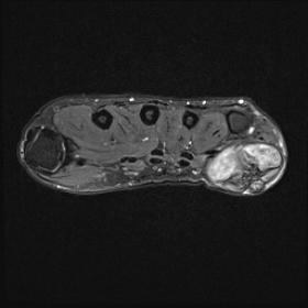
Magnetic resonance imaging (MRI) showed a well-circumscribed lobulated heterogeneous soft tissue mass within the subcutaneous tissues (Fig.1a-d). A thin layer of fat is seen separating the mass from the underlying deep tissues. The deep aspect of the mass demonstrated intermediate signal intensity on T1 weighted imaging and intermediate to high signal intensity on STIR weighted imaging. Superficially within the mass were foci of low signal intensity on T1 and STIR weighted imaging. Computed tomography (CT) images (Fig. 1e+f) showed a soft tissue mass with calcification superficially corresponding to the low T1 and STIR signal intensity on MRI.
Discussion
Ossifying fibromyxoid tumour (OFMT) is a rare soft tissue tumour first described by Enzinger et al. in 1989 [1]. The most common location is within the subcutaneous tissues of the extremities but less commonly is found to involve the trunk and head and neck region. OFMT is classified as a neoplasm of uncertain origin but had previously been suggested to possibly be nerve sheath, myoepithelial or cartilaginous in origin [2].
Clinical presentation of OFMT is usually with a painless well-circumscribed subcutaneous lump measuring around 4-5cm. There is a slight male predilection with median age of approximately 50 years old [3].
CT imaging of OFMTs typically shows a subcutaneous mass with peripheral intralesional ossification. This ossification corresponds to areas of low signal intensity on both T1 and T2 weighted MR images. The myxofibrous component of the tumour demonstrates intermediate signal on T1 weighted imaging and intermediate to high signal on T2 weighted imaging. Occasionally areas of internal haemorrhage are seen which are seen as high signal intensity on both T1 and T2 weighted imaging [4].
OFMT are benign tumours however it has been found that a subset shows atypical histopathological features which correspond to them exhibiting more aggressive behaviour with higher local recurrence and metastatic rates [5]. Core biopsy was performed on this tumour which showed a bland spindle cell tumour, difficult to classify but no evidence of morphological features associated with malignant behaviour. A review of the previously excised specimen from this site was carried out which found features consistent with an ossifying fibromyxoid tumour. In view of these new findings, the core biopsy was felt to likely represent OFMT recurrence. The patient proceeded to surgical excision with the specimen confirmed to be in keeping with OFMT.
This lesion showed no atypical histopathological features so is quoted to have local recurrence rates of 0-12% and metastatic rates of 0-4% [6,7]. The histopathological differential diagnosis includes malignant peripheral nerve sheath tumours which would explain the histological diagnosis of the original tumour in this patient excised 17 years ago [3].
Management of these lesions is usually with complete surgical excision with clinical follow-up, especially if histopathological malignant features are identified.
This case has shown the typical radiological features of a OFMT, a rare soft tissue tumour. It has shown that local recurrence of these tumours can occur even after long time intervals, in this case 17 years. Although OFMT is a rare tumour, it should be considered in the differential of a painless well-defined slow-growing subcutaneous soft tissue mass with peripheral calcification.
Written informed patient consent for publication has been obtained.
Differential Diagnosis List
Final Diagnosis
Ossifying fibromyxoid tumour
Liscense
This work is licensed under a Creative Commons Attribution-NonCommercial-ShareAlike 4.0 International License.
Figures


Medical Imaging Analysis Report
1. Imaging Findings
From the MRI and CT images, a soft tissue mass can be seen in the palmar subcutaneous tissue area near the proximal part of the fifth metacarpal bone of the patient’s right hand. The lesion has a relatively well-defined border, with local ring-like or irregular high-density or calcified areas:
- CT findings: Obvious ossification/calcification density can be seen at the edges or inside of the tumor, presenting as high-density areas that are clearly demarcated from the surrounding subcutaneous soft tissue.
- MRI findings: On T1-weighted images, the tumor mostly shows an intermediate signal, and on T2-weighted images, it may show intermediate to high signal. The calcified/ossified areas appear as low signal on both T1 and T2, consistent with sclerosis or ossification. A small amount of high-signal areas within the mass might indicate mild liquefaction or hemorrhage.
- Location relationship: The tumor is located in the palmar subcutaneous tissue of the fifth metacarpal, with no obvious involvement of adjacent tendons or bone erosion. No clear evidence of abnormal infiltration into the surrounding soft tissue.
2. Potential Diagnoses
Considering the patient’s past medical history (surgical resection in the same location 17 years ago, with pathological findings suggesting a tumor of nerve sheath origin), as well as the recurrence of a slowly growing mass in a similar area and the imaging evidence of ossification/calcification, the following differential diagnoses should be considered:
- Recurrent or relapsed nerve sheath tumor (e.g., schwannoma, malignant peripheral nerve sheath tumor): Schwannomas can present as well-demarcated solid masses, sometimes with degeneration or cystic changes. Although rare, malignant peripheral nerve sheath tumors may show calcification.
- Ossifying Fibromyxoid Tumor (OFMT): This type of rare soft tissue tumor can have a clear sclerotic or ossified rim, commonly found in subcutaneous tissues of the extremities. Both the imaging and pathological features align with those seen in this case.
- Other calcified soft tissue tumors: Such as chondrosarcoma with soft tissue extension, myxoid liposarcoma, etc., but these often involve more prominent soft tissue destruction or distinctive fat signals, which are not strongly indicated in this case.
Overall, given the slow growth, superficial location, distinct ossification/calcification rim, and the patient’s history, OFMT has a higher priority in the differential diagnosis.
3. Final Diagnosis
Taking into account the findings of a review of the patient’s previous pathology, which are consistent with Ossifying Fibromyxoid Tumor (OFMT), and the corresponding imaging features, the most likely diagnosis in this case is:
Recurrence of Ossifying Fibromyxoid Tumor (OFMT).
If there are still doubts or if the histopathological findings suggest any malignant features, a complete surgical excision with further pathological assessment is recommended. Immunohistochemistry and molecular testing may be performed as needed to clarify the benign or malignant nature.
4. Treatment Plan and Rehabilitation
4.1 Treatment Strategy
- Surgical treatment: Complete surgical excision is the mainstay treatment for this type of tumor, aiming to ensure clear margins and reduce local recurrence. If pathology confirms malignancy or unclear surgical margins, re-operation or adjuvant radiotherapy may be considered.
- Follow-up observation: As OFMT can recur and very rarely metastasize, regular imaging follow-up (MRI or ultrasound) is required to monitor for postoperative recurrence.
4.2 Rehabilitation and Exercise Prescription
Given the lesion is located in the palm of the hand, postoperative rehabilitation should focus on restoring hand function once the wound has healed. The following principles are suggested, referring to the FITT-VP (Frequency, Intensity, Time, Type, Progression, Volume) framework:
- Early postoperative phase (1-2 weeks):
- Protective immobilization to minimize grasping, squeezing, or other stressful hand movements;
- Perform passive or assisted active exercises with limited range and gentle force to avoid painful stretching.
- Mid-postoperative phase (2-6 weeks):
- After suture removal and good wound healing, gradually introduce simple grasping and pinching exercises, such as lightly squeezing a soft grip ball or ring;
- Incrementally increase the strength level; each session should not cause significant pain, with 1-2 sessions per day, each lasting 10-15 minutes.
- Late postoperative recovery phase (6 weeks and beyond):
- Incorporate more refined hand function training, such as pinching small objects or writing;
- Depending on overall recovery, gradually resume normal hand activities, including daily gripping tasks and light resistance training.
- Precautions:
- Seek immediate medical consultation if swelling, significant pain, or functional decline occurs;
- Avoid heavy impact or excessive force training, particularly around the ossified area or surgical site.
5. Disclaimer
This report is based solely on the provided imaging and medical history for a preliminary analysis and does not replace an in-person clinical evaluation. Please consult a professional attending physician or specialized medical team for final decisions on diagnosis and treatment. If any questions arise or new symptoms develop, seek medical attention promptly.
Human Doctor Final Diagnosis
Ossifying fibromyxoid tumour