Calcaneal Osteosarcoma


Clinical History
A 53-year-old man presented to an orthopaedic department with a history of left ankle pain over the past 3 weeks with swelling that worsened with activity and in the morning. No trauma was reported. The laboratory markers of inflammation were not elevated. At physical examination moderate swelling of the ankle without notable impact on range of motion was observed.
Imaging Findings
No pathological findings were observed on X-ray.
CT of the left ankle showed an ill-defined hypodense lesion in the calcaneum with no cortical destruction or periosteal reaction. No calcification or signs of mineralization was observed.
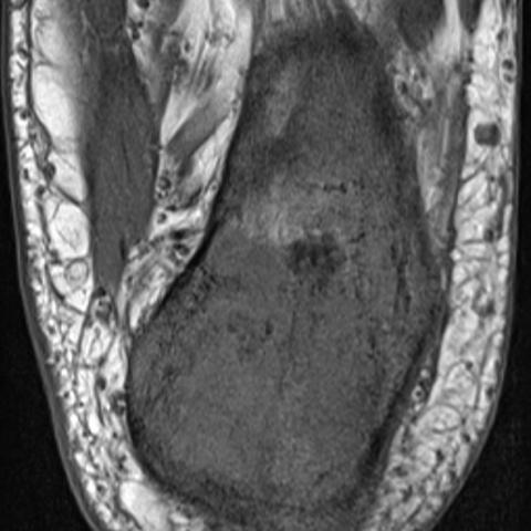
MRI revealed a diffuse hyperintense signal on fat-suppressed proton density-weighted image and diffuse hypointense signal on T1-weighted image of the calcaneus. After contrast administration, heterogeneous enhancement of the calcaneus was observed. There was no evidence of periosteal reaction or extraosseous expansion. Mild oedema of the surrounding soft tissues without ankle joint effusion were observed.
Scintigraphic imaging confirmed the increased uptake of the radioactive substance of the left calcaneus. No signs of tumour spread or metastasis was observed on bone scintigraphy. Thoracic metastases were excluded with additional thoracic CT imaging.
Discussion
Osteosarcoma is the most common non-haemopoietic primary malignant neoplasm of bone. It typically affects people from 10 to 30 years old, with a peak incidence in the second decade of life, showing slight male predominance [1, 2]. The diaphysis of the long bones are the most common sites where osteosarcoma may arise [1,3]. Osteosarcoma of the foot accounts only for 1% of all osteosarcoma cases [4]. Within the foot it arises from the calcaneus or metatarsal bones in 75% [4]. These cases are more likely to be low grade and have higher reported survival rate [4].
Symptoms of osteosarcoma presentation are non-specific. Progressively worsening pain and swelling in the affected ankle joint are the major complaints. These were observed in our case. Physical examination typically reveals tender swelling and restricted joint mobility [1,4]. Even though NSAID administration causes good initial improvement of symptoms, they are often the cause for delayed diagnosis and treatment [4].
Typical radiograph and CT appearance of the calcaneal osteosarcoma can be very informative, including dense sclerotic, lytic lesion, cortical destruction and extraosseous expansion [1, 5]. In our case no lesion was detected on radiograph, however, CT showed a lytic lesion with no cortical destruction, periosteal reaction, calcification or signs of mineralization. MRI should be performed for preoperative staging and offers good visualisation of extraosseal infiltration [1]. MRI signal of the lesion is usually heterogeneous due to mixture of lysis, sclerosis and rarefaction [1]. Heterogeneous signal on MRI was also observed in our case, on account of necrotic and sclerotic lesions in combination with areas of preserved bone structure. MRI can help in differentiating the tumour, but only histopathology offers definitive diagnosis [1]. Our histopathology exam revealed malignant sarcomatous cells arranged in cords and nests with associated osteoid deposition. Bone scintigraphy is performed in order to rule out osseous metastases [6]. Additionally, CT of the chest ought to be performed to rule out metastatic disease at the time of diagnosis [1, 4].
The treatment of the bone sarcomas varies depending on the stage. Standard treatment of patients with the osteosarcoma consists of a combination of chemotherapy and surgery [4,7]. Limb-salvage surgery of the foot osteosarcoma is usually not possible, due to poor compartmentalisation of the foot. Therefore, as in our case, below the knee amputation is surgery of choice [4]. Currently, the 5-year survival rate of all osteosarcomas after adequate therapy is approximately 60-80%, depending on the tumour’s response to neoadjuvant chemotherapy [1,7].
Teaching points: Although osteosarcoma is one the most common primary malignant tumours of bone, it rarely occurs in the feet. It can be detected on radiograph or CT, but MRI must be performed for preoperative staging. The final diagnosis is made by histopathology examination. Treatment usually consists of a combination of chemotherapy and surgery.
Written informed patient consent for publication has been obtained.
Differential Diagnosis List
Final Diagnosis
Calcaneal osteosarcoma
Liscense
This work is licensed under a Creative Commons Attribution-NonCommercial-ShareAlike 4.0 International License.
Figures


1. Imaging Findings
Based on the provided X-ray, CT, and MRI images of the left ankle, the following observations can be made:
- In the calcaneus region, there is localized bone destruction on imaging. On CT, it appears as a lytic lesion, but overall the cortex remains intact, with no obvious cortical disruption, periosteal reaction, or significant soft tissue swelling.
- On MRI, the lesion shows heterogeneous signal intensity, with areas of low and high signal interlaced, suggesting the possibility of necrosis, sclerosis, and a small amount of normal bone mixed in.
- Mild soft tissue reaction can be seen around the lesion, but there is no evidence of a significant soft tissue mass breaking through or extensive infiltration.
2. Potential Diagnoses
Taking into account the patient’s age (53 years), clinical history (no trauma, recent progressive pain and swelling), and the imaging findings (lytic lesion, heterogeneous signals, mild local tissue involvement, etc.), the following differential diagnoses should be considered:
- Osteosarcoma:
- Clinically, it is one of the most common primary malignant bone tumors, typically seen in individuals aged 10–30, but it can also occur in older adults.
- Typical imaging features often include mixed lytic or sclerotic changes, possible periosteal reactions, and soft tissue masses.
- Osteosarcoma in the foot is rare, especially in the calcaneus. It often presents as a lytic lesion, and some patients may lack a typical periosteal reaction, leading to misdiagnosis or missed diagnosis.
- Giant Cell Tumor of Bone:
- More common in adults aged 20–40, often found in the epiphysis or metaphysis, and can present as a lytic lesion.
- Commonly involves areas around the knee joint or distal radius; it is less frequent in the foot, though the calcaneus can be affected.
- Imaging can show a “soap-bubble” appearance with well-defined margins. When MRI reveals soft tissue components, it may indicate aggressiveness.
- Chondrosarcoma:
- Often occurs in middle-aged and older adults, typically at a later onset compared to osteosarcoma.
- Imaging might show lytic or calcified areas, with characteristic “rings and arcs” calcification in the cartilaginous matrix.
- In this case, CT does not show significant calcification or mineralization, so chondrosarcoma is relatively less likely.
3. Final Diagnosis
Considering the patient’s age, clinical presentation and symptoms (pain and swelling), imaging findings (a lytic lesion in the calcaneus with no obvious cortical destruction or periosteal reaction, but with MRI indicating a rather extensive lesion), and pathological examination (malignant sarcoma cells with osteoid deposition), the most likely diagnosis is:
Osteosarcoma
This diagnosis has been confirmed by histopathological results, indicating an osteosarcoma of the calcaneus.
4. Treatment Plan and Rehabilitation
Treatment for osteosarcoma generally involves a multidisciplinary approach:
- Surgical Treatment: Given the lesion’s extent and the unique anatomy of the foot, limb-sparing procedures can be challenging for foot osteosarcomas. In this case, with the tumor in the calcaneus and confirmed malignancy, surgical excision is indicated. Some patients may require below-knee amputation to ensure complete tumor resection and improve long-term survival.
- Chemotherapy: Standard treatment includes neoadjuvant (preoperative) and adjuvant (postoperative) chemotherapy regimens targeting osteosarcoma, to control tumor spread or reduce the risk of recurrence.
- Postoperative Rehabilitation: For patients requiring amputation, postoperative care includes prosthetic fitting and rehabilitation training to gradually restore walking function.
Rehabilitation and Exercise Prescription (FITT-VP):
- Frequency: Begin with rehabilitation exercises 1–2 times per day, adjusting as tolerated.
- Intensity: Start with low-intensity exercises (such as muscle strengthening and range-of-motion exercises), avoiding excessive weight-bearing, especially in the early postoperative period, to focus on wound healing and residual limb pain management.
- Time: Each session should last around 15–20 minutes initially, gradually increasing to 30 minutes or longer, provided there is no significant discomfort.
- Type:
- Early stages can include active and passive hip and knee range-of-motion exercises in sitting or supine positions.
- Later stages include prosthetic training, working on standing balance, slow walking, and activities of daily living.
- Under professional supervision, residual limb strengthening and core stability exercises can be introduced.
- Progression: As the patient adapts, gradually increase the range of motion and weight-bearing based on age, cardiovascular capacity, and surgical recovery, ensuring a steady escalation in exercise difficulty.
- Volume & Progression:
- Track total training volume (e.g., total weekly exercise time) or weight-bearing intensity to measure progress.
- Increase training volume gradually under safe conditions, while closely monitoring pain, wound healing, and gait stability.
Throughout the process, closely monitor the skin condition of the residual limb to prevent ulcers or pressure injuries from prosthetic use.
Disclaimer
This report is based on current imaging and clinical information for preliminary analysis and recommendations, for reference only. It is not a substitute for in-person consultation or the advice of professional physicians. Any treatment or rehabilitation regimen should be carried out under the guidance of qualified medical personnel.
Human Doctor Final Diagnosis
Calcaneal osteosarcoma